Clavicle fractures are common after a fall onto the shoulder, especially in children. The bone is superficial along its whole length, so a linear probe shows its cortex clearly. In children, studies have found ultrasound accurate for clavicle fractures, and it can be done without moving a painful shoulder.
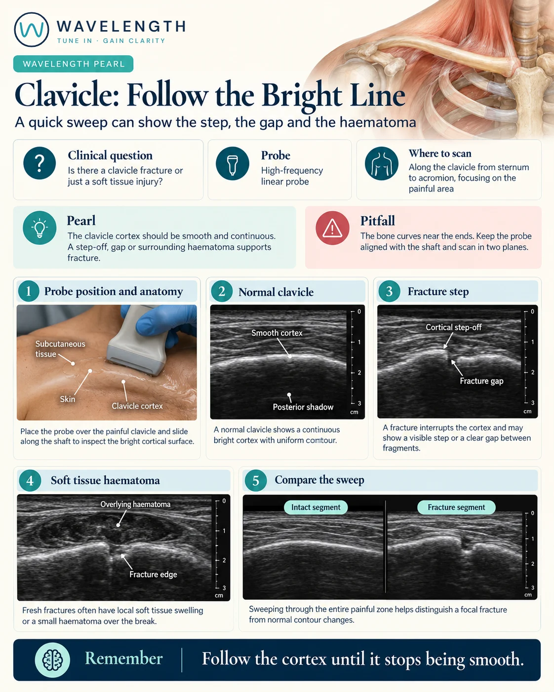
Scan images in the graphic are illustrations, not patient scans.
The scan
- Sit the patient up, supported, with the arm resting.
- Place a high-frequency linear probe along the clavicle, so the cortex is a bright curved line.
- Sweep from the sternal end to the acromion, keeping the probe on the line of the bone as it curves.
- Turn to short axis over any abnormality.
- Compare with the other side if you are unsure.
Reading the view
- Normal clavicle: a smooth, continuous bright line with a shadow beneath.
- Fracture: a step, a gap or overlapping ends, most often in the middle third.
- Supporting signs: a haematoma over the bone, and tenderness as the probe passes.
- The ends: the acromioclavicular joint at the shoulder, and the sternoclavicular joint in the middle. A step here may be a joint injury rather than a fracture.
Action
Most midshaft fractures are treated in a sling through your fracture pathway, with an X-ray if your pathway requires one. Skin tented or broken over the fracture, a cold or weak arm, or a posterior sternoclavicular dislocation (pain at the sternal end, difficulty swallowing or breathing) needs urgent orthopaedic review; the last needs CT.