Distal triceps rupture is uncommon and often missed. It follows a fall onto an outstretched hand, a direct blow, or heavy lifting, and is more likely with steroid use or kidney disease. Patients may still extend the elbow with gravity, so the weakness is easy to overlook. Like the distal biceps, the tendon is superficial and easy to follow to its insertion.
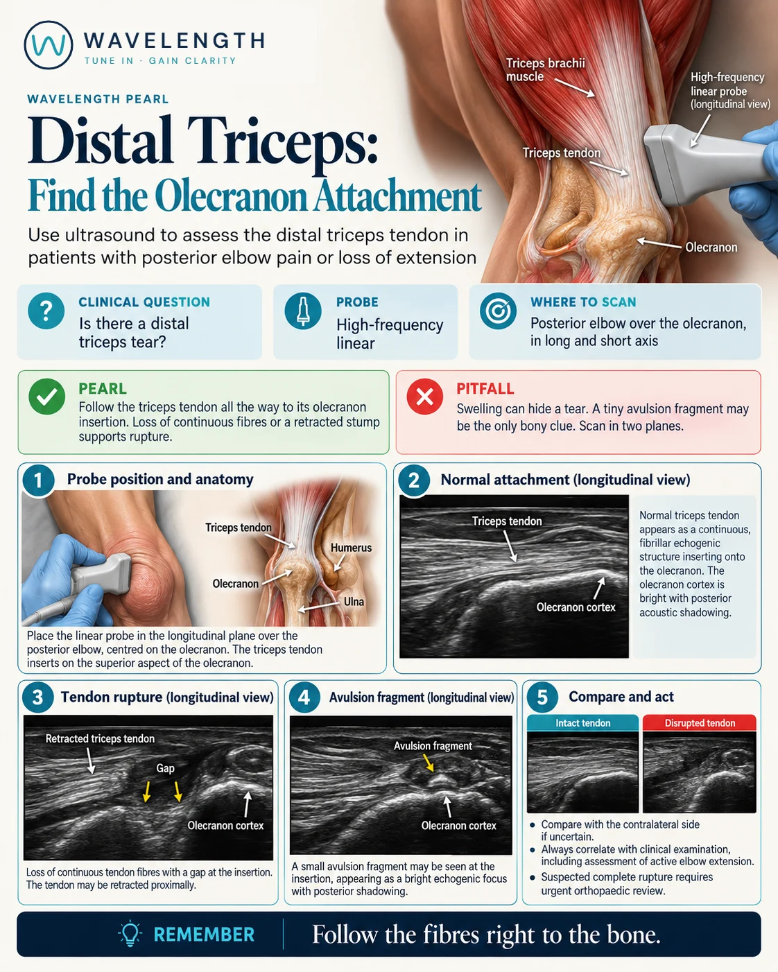
Scan images in the graphic are illustrations, not patient scans.
The scan
- Rest the arm across the chest or on a pillow with the elbow bent, so the back of the elbow faces you.
- Use a high-frequency linear probe in long axis over the back of the elbow, centred on the olecranon.
- Follow the triceps tendon from the muscle down to its insertion on the olecranon.
- Turn to short axis and sweep across the tendon.
- Gently flex and extend the elbow while you watch, and compare with the other side.
Reading the view
- Normal: a bright, fibrillar tendon inserting onto the bright olecranon cortex.
- Complete tear: a dark gap at the insertion, with the tendon end pulled back up the arm.
- Avulsion: a small bright fragment with a shadow, lying above the olecranon within the tendon end. The same flake may show on a lateral X-ray.
- Partial tear: some intact fibres, often the deep or lateral part, with a dark defect.
Action
Test extension against gravity and against resistance. Suspected complete rupture needs urgent orthopaedic review, because early repair gives the best results. A lateral elbow X-ray looks for the avulsion flake and other injuries.