Infection inside the flexor tendon sheath spreads quickly along the finger and into the palm, and can destroy the tendon. The classic Kanavel signs are a finger held slightly bent, swelling of the whole finger, tenderness along the sheath, and pain on passive straightening. Ultrasound adds a look inside the sheath, and shows whether a collection is in the sheath or above it.
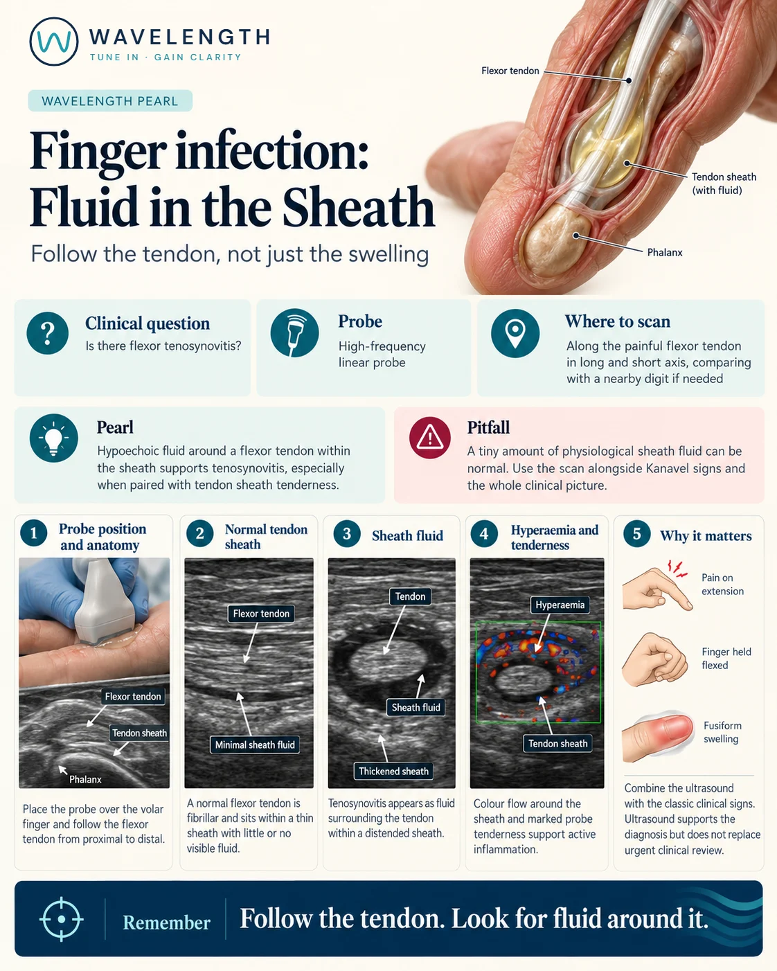
Scan images in the graphic are illustrations, not patient scans.
The scan
- Rest the hand palm up. Use a high-frequency linear probe with plenty of gel and very little pressure.
- Find the flexor tendon on the front of the finger, a bright fibrillar band over the phalanges.
- Follow it from the fingertip into the palm in long axis, then in short axis.
- Look for fluid around the tendon, and its thickness and texture.
- Add colour Doppler around the sheath.
- Compare with the same tendon in a neighbouring finger.
Reading the view
- Normal: a bright tendon in a thin sheath, with little or no fluid around it.
- Tenosynovitis: fluid surrounding the tendon within a distended, often thickened sheath, with increased flow around it and tenderness under the probe.
- Superficial collection: fluid above the sheath, under the skin, with the tendon and sheath normal beneath it.
- Cellulitis: swollen, cobblestoned tissue without fluid around the tendon.
Action
Suspected flexor tenosynovitis needs urgent hand surgery review, intravenous antibiotics, elevation and splinting, whatever the scan shows; many patients need surgical washout. Ask about penetrating injuries and bites, which are high risk.