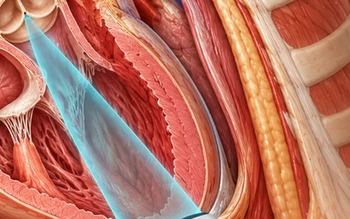
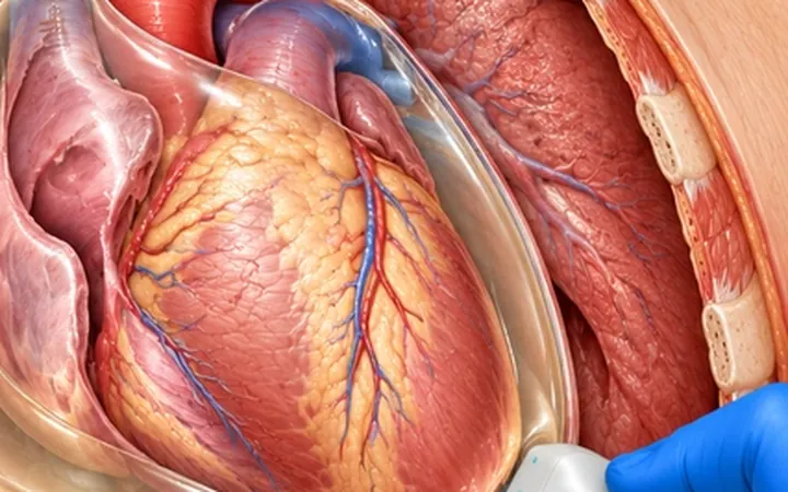
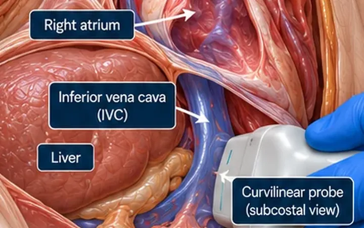
A plethoric IVC that does not collapse with breathing usually points to raised right atrial pressure, from fluid overload, right heart failure, tamponade or a large pulmonary embolism. Occasionally the vein is wide because something is inside it. Thrombus can extend up from the leg and pelvic veins or form around a femoral line or IVC filter. Tumour can grow into the IVC from the kidney or liver. The view you already take for fluid status can show it.
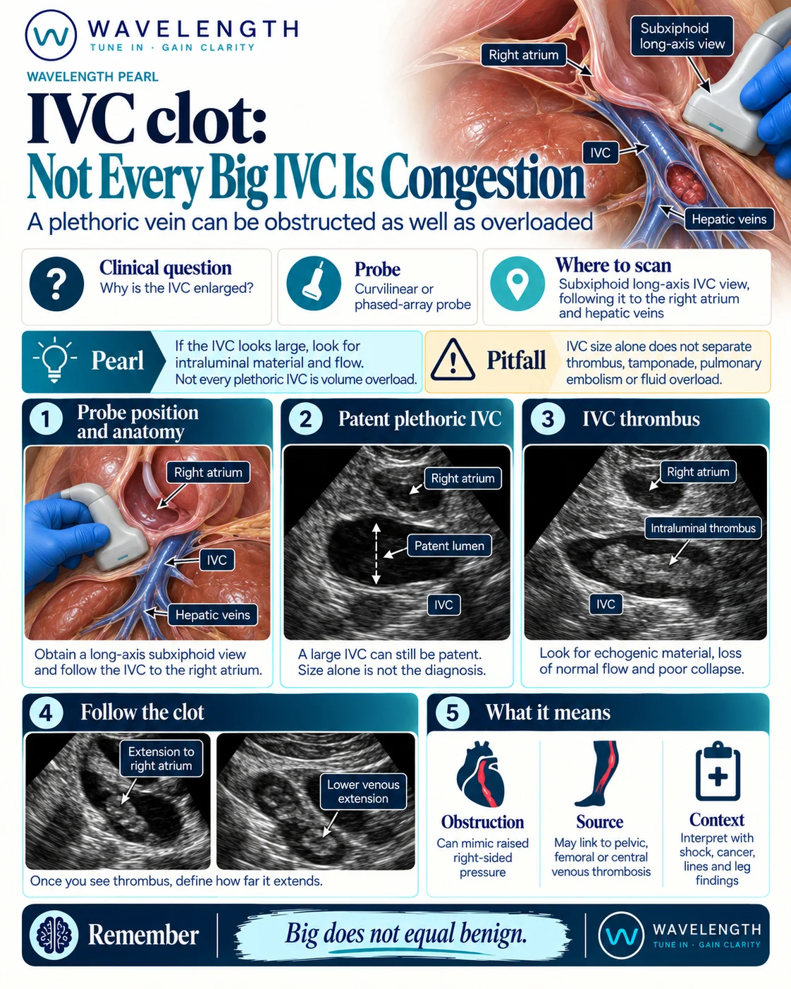
Scan images in the graphic are illustrations, not patient scans.
The scan
- Use the curvilinear or phased-array probe in the subxiphoid window, in long axis.
- Follow the IVC into the right atrium, as in the IVC pearl, and confirm the hepatic veins draining into it.
- Before you measure, look inside the lumen. Increase the gain a little to bring out faint echoes.
- Add colour Doppler with a low scale. Look for flow around or through any material.
- If you see material, sweep up into the right atrium and down towards the renal veins to find both ends.
Reading the view
- Patent plethoric IVC: wide and still, but black inside with flow throughout. Interpret it with the heart and the clinical picture.
- Thrombus: echogenic material within the lumen, with absent or reduced flow around it. Fresh clot can be almost black, so use colour.
- Clot in the right atrium: a mobile mass extending into or sitting within the right atrium, a clot in transit.
- Tumour thrombus: material extending from a renal vein or the liver; it may contain its own blood flow.
Action
Thrombus in the IVC needs senior review, anticoagulation decisions and formal imaging, usually CT venography or contrast CT. A mobile clot in the right atrium is an emergency: involve senior, cardiology and critical care colleagues at once, because of the risk of pulmonary embolism.