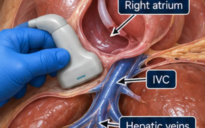
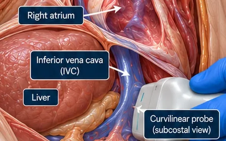
The left ventricular outflow tract velocity time integral (LVOT VTI) is the distance a column of blood travels through the outflow tract in one beat. If the outflow tract stays the same size, a change in VTI reflects a change in stroke volume. Its strength is repetition. Its weakness is comparing two measurements which were taken differently.
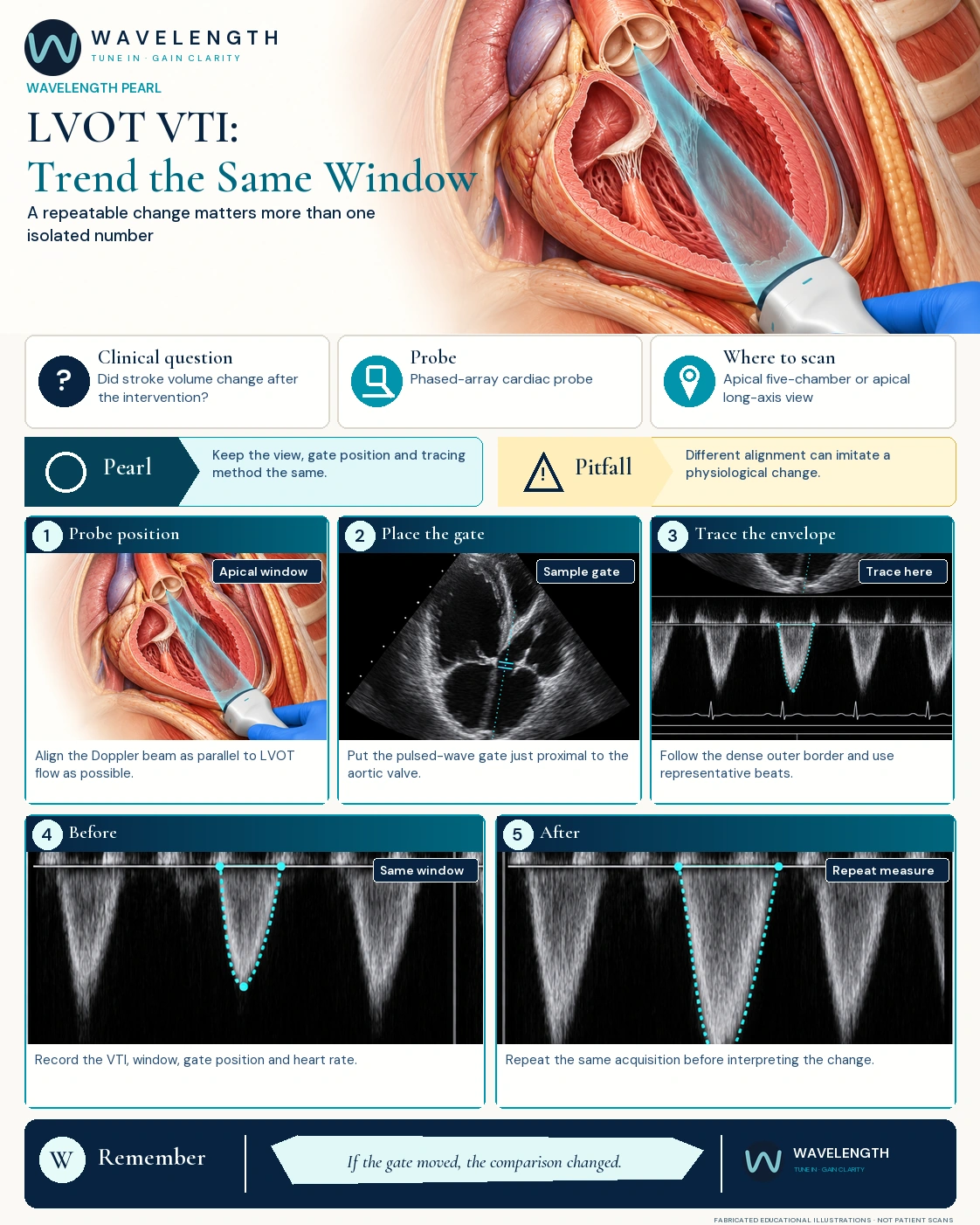
Scan images in the graphic are fabricated educational illustrations, not patient scans.
The scan
- Get the best apical five-chamber or apical long-axis view you can reproduce.
- Line the Doppler beam up with the flow through the outflow tract.
- Place the pulsed-wave gate in the outflow tract, just below the aortic valve, away from the acceleration at the valve itself.
- Record several clean envelopes. Trace the outer edge of the dense part of the signal, the same way every time.
- Average beats: at least three in a regular rhythm, more in an irregular one.
- After a fluid bolus, a passive leg raise or a drug, return to the same window, depth, gate position and tracing method.
Reading the trend
- A clearly larger repeat VTI, taken the same way, supports a rise in stroke volume.
- Record the heart rate too. Cardiac output is stroke volume times heart rate.
- One value is not a fluid prescription. Read it with the circulation, the rhythm, the valves and ventricular function.
Action
Use VTI as one part of a focused haemodynamic reassessment. Write down the window and settings so the next measurement is truly comparable.