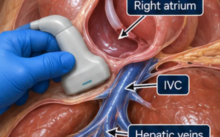
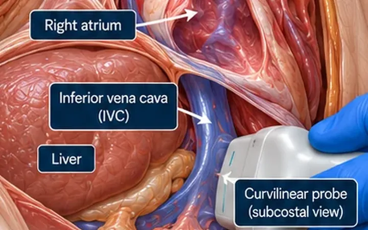
A pericardial effusion raises the question of tamponade, but the size of the effusion does not answer it. Fluid matters when the pressure it creates stops the right heart filling. That is why the timing of right-sided chamber collapse tells you more than the fact that a wall moves inward.
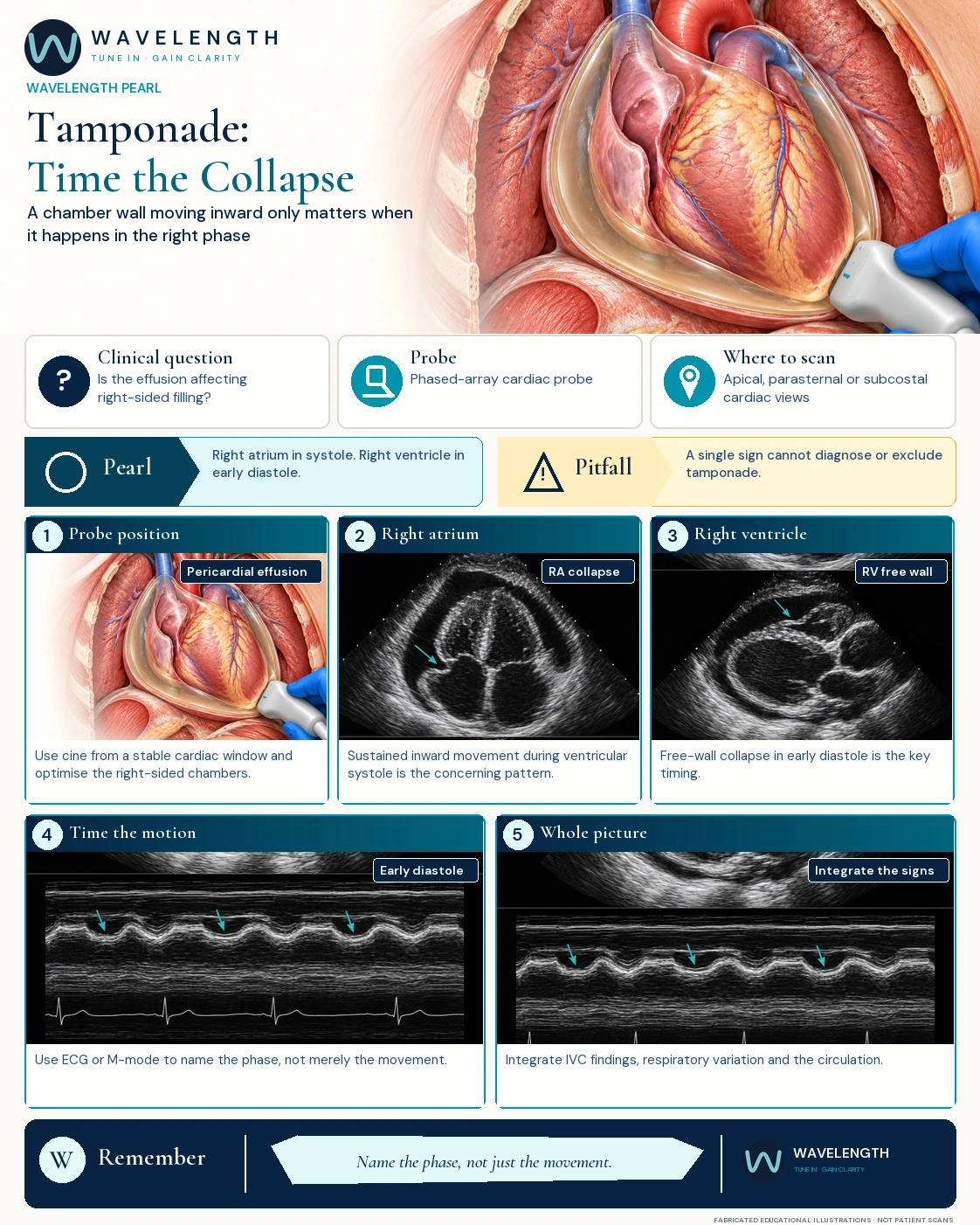
Scan images in the graphic are fabricated educational illustrations, not patient scans.
The scan
- Confirm the effusion in more than one view: parasternal long axis, subcostal and apical four-chamber.
- Record a cine loop, with an ECG trace on the screen if the machine has one.
- In the apical or subcostal four-chamber view, watch the right atrial free wall. Inward movement which lasts through ventricular systole is the concerning pattern.
- In the parasternal or subcostal view, watch the right ventricular free wall. Collapse in early diastole, as the ventricle should be filling, is the concerning pattern.
- Use M-mode through the right ventricular free wall when it helps you time the movement against the mitral valve or the ECG. Do not let a poorly placed cursor replace the cine loop.
- Finish the picture: the IVC and its change with breathing, left and right ventricular function, and the patient's circulation.
Reading the view
- Right atrium: a brief inward flicker is common and unhelpful. Collapse lasting more than about a third of the cardiac cycle is more specific.
- Right ventricle: inward collapse in early diastole is more specific than atrial collapse, and supports tamponade physiology.
- Context changes the signs: a dry patient can collapse at lower pericardial pressure, so the signs appear early. Raised right-sided pressures, from pulmonary hypertension or a stiff right ventricle, can hide them.
- The IVC: a full IVC which barely changes with breathing is sensitive but not specific. An IVC which collapses well makes tamponade unlikely.
Action
A shocked patient with a pericardial effusion and supportive chamber collapse needs urgent senior, cardiology and procedural help through your local pathway. Do not wait to perfect the measurements.