Septic arthritis of the sternoclavicular joint is uncommon, which is why it is missed. Injecting drug use, diabetes, immunosuppression, a central venous line and infection elsewhere are the usual risk factors. The joint is small and superficial, so ultrasound can show an effusion within a minute, and the other side is right next to it for comparison. Like the hip and the wrist, the sign is fluid lifting the capsule away from the bone.
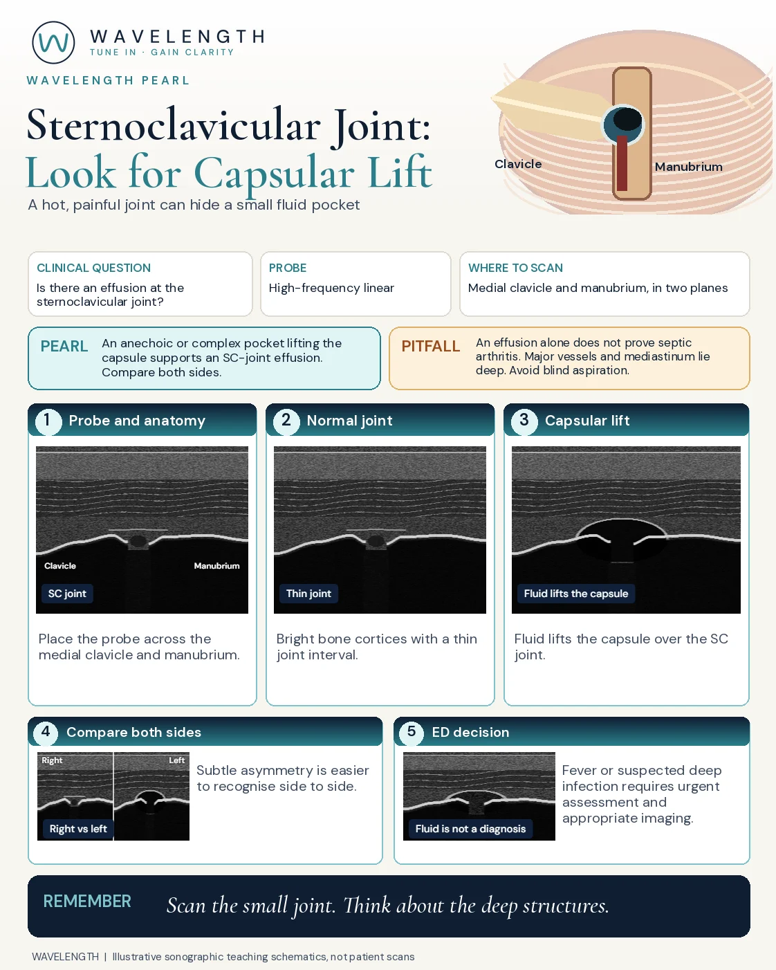
Scan images in the graphic are illustrations, not patient scans.
The scan
- Lie the patient flat with the head straight.
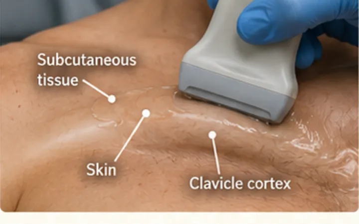
- Use a high-frequency linear probe across the joint: along the medial clavicle, onto the manubrium.
- Find the two bright bone surfaces and the narrow gap between them.
- Turn the probe 90 degrees for a second plane.
- Scan the other joint with the same settings and compare.
Reading the view
- Normal: two bright cortices, the clavicle head and the manubrium, each with a shadow beneath, a narrow dark gap and a thin capsule lying close to the bone.
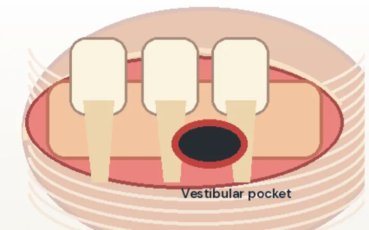
- Effusion: an anechoic or complex pocket that lifts the capsule into a convex bulge over the joint.
- Beyond the joint: fluid tracking into the soft tissue of the chest wall, or an irregular, broken cortex, suggests an abscess or osteomyelitis.
Action
With fever or raised inflammatory markers, take blood cultures and treat as possible septic arthritis. CT or MRI shows deep extension. Aspiration, if needed, is image-guided by an experienced operator, with orthopaedic review.