A swollen wrist is common: after a fall, with gout or pseudogout, with inflammatory arthritis, and rarely with infection. The swelling on the surface does not show which layer it is in. Ultrasound does, in a minute, and it guides the needle if fluid needs to come out.
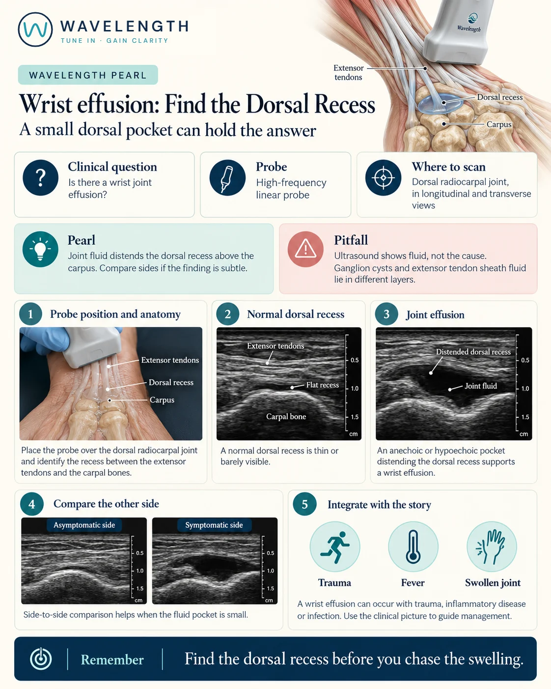
Scan images in the graphic are illustrations, not patient scans.
The scan
- Rest the hand palm down on a pillow, with the wrist slightly flexed.
- Place a high-frequency linear probe in long axis over the back of the wrist, in line with the middle finger.
- Find the distal radius, then the lunate and capitate below it. The dorsal recess lies over these bones, deep to the extensor tendons.
- Scan in short axis across the wrist.
- Compare with the other wrist if the finding is small.
Reading the view
- Normal: a thin or barely visible recess over the carpal bones.
- Effusion: a dark pocket lifting the recess between the bones and the tendons.
- Tenosynovitis: fluid around a tendon, which moves with the tendon when the fingers move.
- Ganglion: a well-defined dark cyst, often with a neck leading towards the joint.
- Cellulitis: swollen, cobblestoned tissue under the skin, with no pocket.
Action
A hot wrist that might be infected needs aspiration, under ultrasound guidance if possible, and the fluid sent for microscopy, culture and crystals before antibiotics. After a fall, follow your scaphoid and wrist fracture pathways.