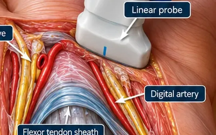
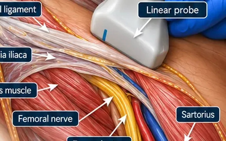
The femoral nerve supplies the front of the thigh, most of the femur, the front of the knee and, through the saphenous nerve, the inner side of the lower leg. A femoral nerve block gives fast analgesia for a femoral shaft fracture, a fractured neck of femur or a painful knee injury, and makes splinting and transfers bearable. In children with a femoral shaft fracture it is often the best first analgesic.
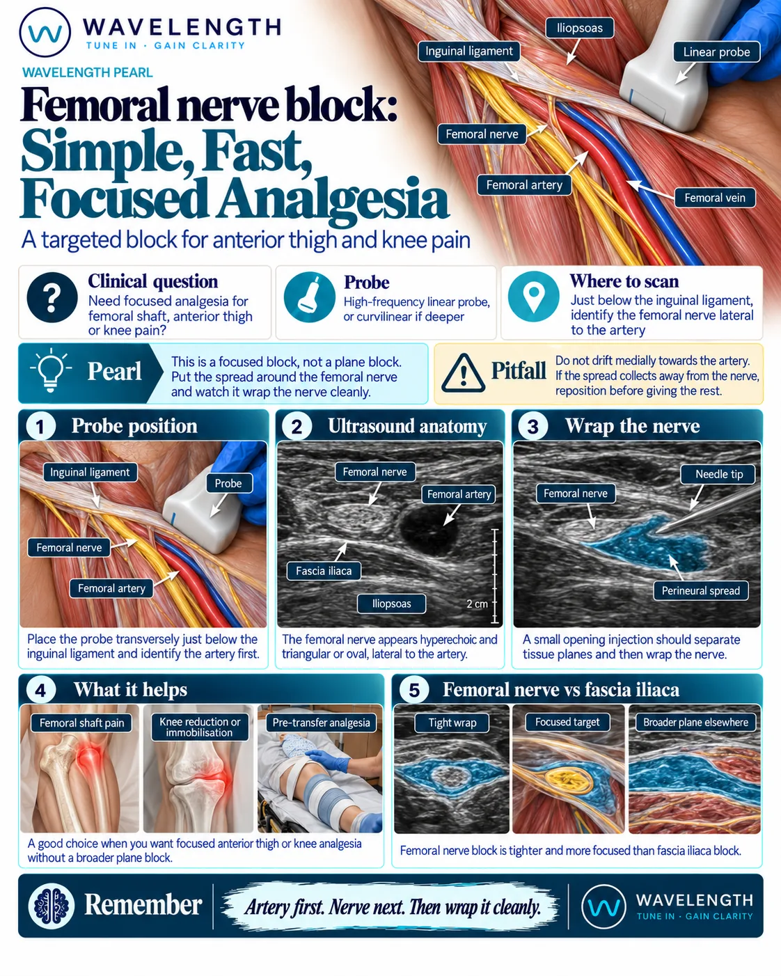
Scan images in the graphic are illustrations, not patient scans.
Before you block
- Record the neurovascular examination of the leg, including the distal pulses.
- Record consent, calculate the maximum safe dose of local anaesthetic, attach monitoring and know where the lipid emulsion is.
- Think about compartment syndrome. In a high-risk injury, such as a tibial fracture, agree the plan for monitoring with the orthopaedic team first.
The scan
- Lie the patient supine with the leg straight.
- Place the linear probe transversely just below the inguinal ligament, over the femoral pulse.
- Find the femoral artery first: a round, black, pulsating circle. The femoral vein lies medial to it and collapses with pressure.
- Look lateral to the artery for the nerve: a bright, flattened oval or triangle, lying on iliopsoas and under the bright line of fascia iliaca.
- Insert the needle in plane from the lateral side, so the needle meets the nerve before it reaches the artery.
- Pass through fascia iliaca, inject 1 to 2 mL, and watch the fluid lift the fascia and spread around the nerve. Then inject the rest, usually 10 to 20 mL.
Reading the view
- Femoral artery. Round, black and pulsating. Colour Doppler confirms it.
- Femoral nerve. Bright and honeycomb in texture, lateral to the artery, deep to fascia iliaca.
- Correct spread. Dark fluid under fascia iliaca, wrapping above and below the nerve.
- Wrong layer. Fluid above fascia iliaca spreads in the fat and does not reach the nerve.