Nerve blocks

Digital nerve block: block the base, one deposit on each side

A digital nerve block numbs a finger for a laceration, a nail bed repair or a reduction. The palmar digital nerves run on each side of the flexor tendon sheath. Place a small deposit beside each one, and use ultrasound to see them.

Each finger has two palmar digital nerves, running beside the flexor tendon sheath with the digital arteries, and two smaller dorsal digital nerves. The palmar nerves supply the palmar surface and, in the index, middle and ring fingers, the back of the finger beyond the middle joint, including the nail bed. Many clinicians perform digital blocks by landmark. Ultrasound shows the nerves and arteries, and confirms the spread with a smaller volume.
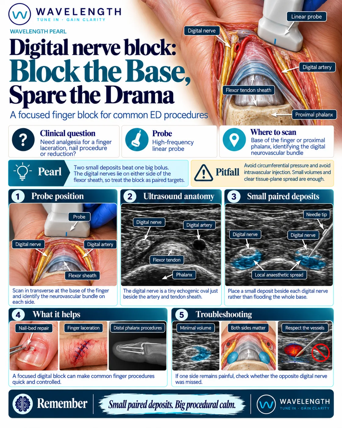
Wavelength Pearl: digital nerve block. Place a small deposit beside each digital nerve at the base of the finger.

Scan images in the graphic are illustrations, not patient scans.

Before you block

  1. Record sensation on both sides of the finger and the tendon function before the block. A digital nerve injury must be documented first.
  2. Record consent. One to two millilitres on each side is usually enough.
  3. Lidocaine with adrenaline is safe in the fingers of healthy patients. A large multicentre study found no finger loss. Follow your trust's guidance, and avoid adrenaline where the circulation to the finger is already poor.

The scan

  1. Rest the hand palm up. Use plenty of gel, or a gel pad, over the base of the finger.
  2. Place a high-frequency linear probe transversely over the palmar surface of the proximal phalanx.
  3. Identify the bone, the bright flexor tendon in its sheath, and on each side the small digital artery with the nerve beside it. Colour Doppler finds the arteries.
  4. Insert the needle from the side of the finger, in plane or out of plane, and place a small deposit beside the nerve on one side.
  5. Repeat on the other side.

Reading the view

  • Proximal phalanx. A curved bright line with a shadow below.
  • Flexor tendon. A bright, fibrillar oval on the bone.
  • Digital artery. A small black circle on each side, with flow on Doppler.
  • Digital nerve. A tiny bright oval beside each artery.
  • Correct spread. A small pool of fluid around each nerve.
Test yourself

5 questions on this pearl

Answer at your own pace. Each answer comes with a short explanation. About 3 minutes.

Take the test

Practise it with us

Four or five delegates per instructor, FAMUS-accredited faculty and long, supervised time on the probe.

See the core course

More on nerve blocks

Nerve blocks

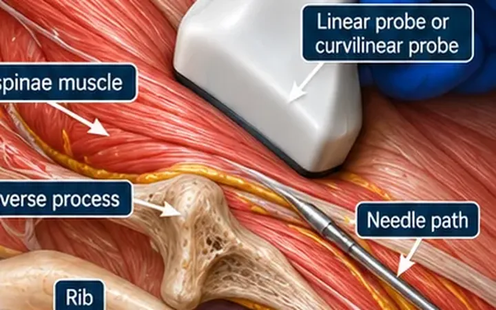
Erector spinae plane block: bone, muscle, then flood the plane

The erector spinae plane block places local anaesthetic on the transverse process, under erector spinae, far from the pleura. Find the flat-topped transverse process, lift the muscle off it and watch the fluid spread up and down.

2 min read · 5 questions

Nerve blocks

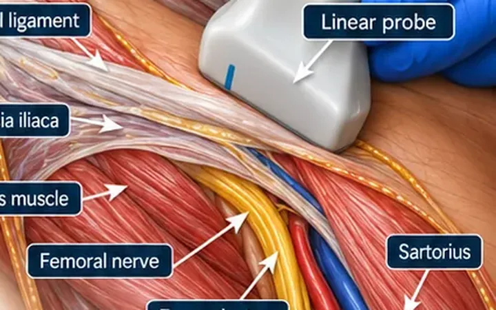
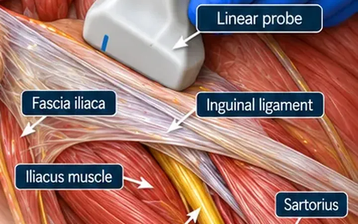
Fascia iliaca block, infra-inguinal: find the fascia, then flood the plane

The classic emergency department block for a fractured hip. Find fascia iliaca lying on iliacus, lateral to the femoral nerve, and fill the plane with a larger volume so it spreads across to the nerves of the thigh.

2 min read · 5 questions

Nerve blocks

Fascia iliaca block, supra-inguinal: go above the ligament for better spread

The supra-inguinal approach places the local anaesthetic under fascia iliaca above the inguinal ligament, where it spreads up towards the lumbar plexus and reaches the lateral femoral cutaneous nerve more reliably. Find iliacus in a sagittal view near the anterior superior iliac spine and watch the fluid travel towards the head.

2 min read · 5 questions

All nerve blocks pearls

Newsletter

Get the next one by email.

One email a week: a practical scanning skill, new Learn posts and course dates before they go public.