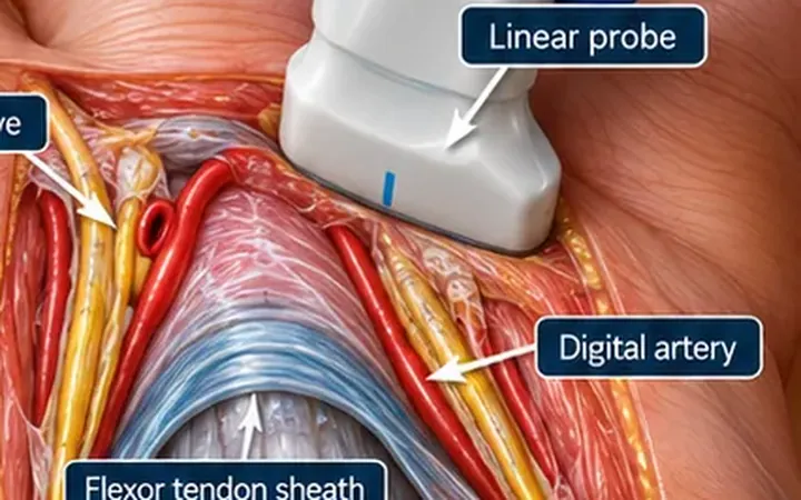
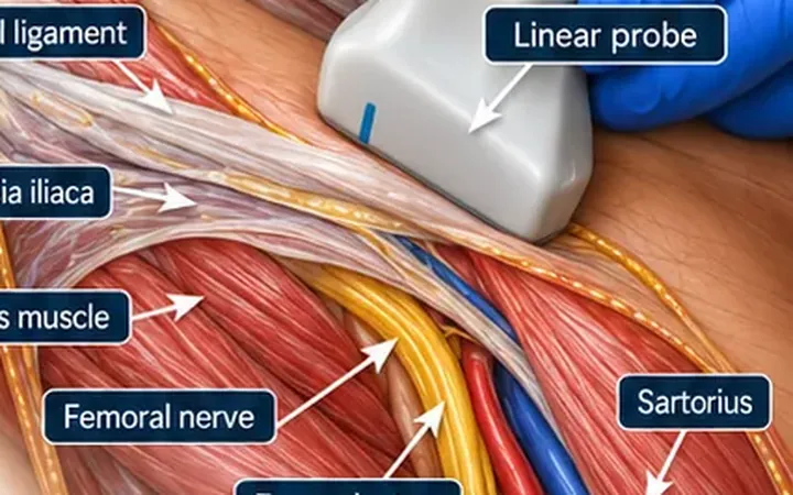
The supra-inguinal fascia iliaca block was described in 2011 as a way to place the injection higher, inside the pelvis, under the same fascia. Cadaver and clinical studies show more consistent spread to the femoral and lateral femoral cutaneous nerves than the infra-inguinal approach, and it reduces opioid use after hip surgery. In the emergency department it suits a fractured neck of femur or a painful pubic ramus fracture, under local governance.
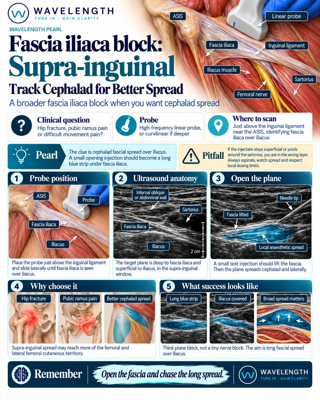
Scan images in the graphic are illustrations, not patient scans.
Before you block
- Record the neurovascular examination and consent, following your trust's process for a patient who lacks capacity.
- Calculate the maximum safe dose. This block uses 30 to 40 mL.
- Attach monitoring and know where the lipid emulsion is.
The scan
- Lie the patient supine.
- Place the linear probe in a sagittal plane, medial to the anterior superior iliac spine, just above the inguinal ligament.
- Identify iliacus lying in the iliac fossa, the bright line of fascia iliaca on top of it, and the abdominal wall muscles above. Sartorius sits superficial at the lower end of the image. The muscles above the fascia form a bow tie shape.
- Colour Doppler finds the deep circumflex iliac artery, which runs just above the fascia and marks the level of the plane.
- Insert the needle in plane from the lower end of the probe, aiming upwards under fascia iliaca.
- Inject 2 to 3 mL and watch the fluid lift the fascia and run towards the head along iliacus. Then inject the rest.
Reading the view
- Iliacus. A thick, dark muscle sloping into the pelvis.
- Fascia iliaca. The bright line on top of iliacus.
- Correct spread. A long strip of fluid under the fascia, running towards the head.
- Wrong layer. Fluid among the abdominal wall muscles or around sartorius.