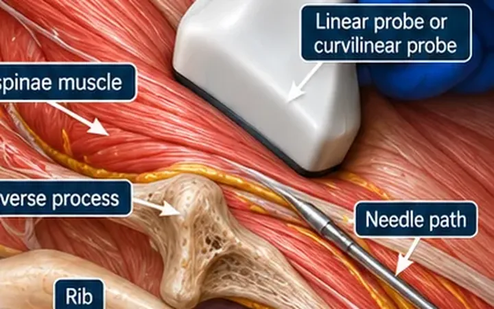
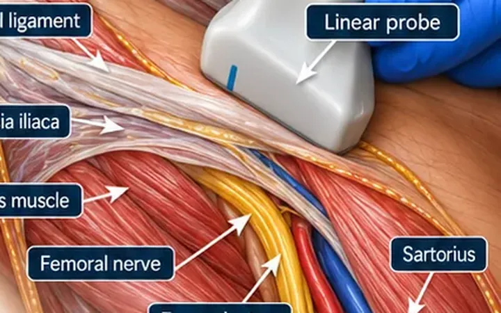
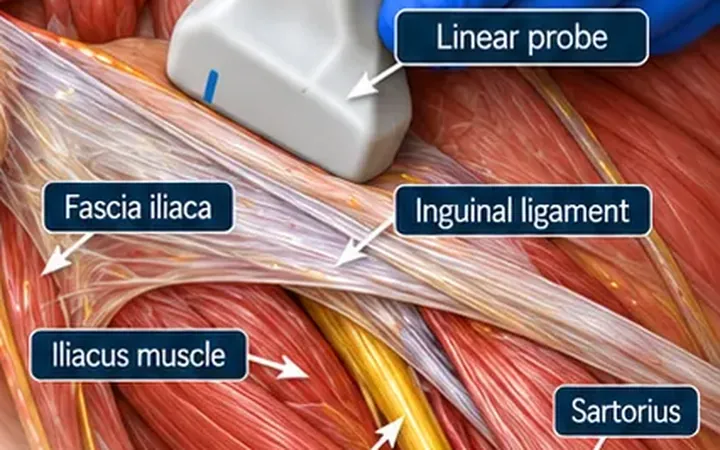
Each finger has two palmar digital nerves, running beside the flexor tendon sheath with the digital arteries, and two smaller dorsal digital nerves. The palmar nerves supply the palmar surface and, in the index, middle and ring fingers, the back of the finger beyond the middle joint, including the nail bed. Many clinicians perform digital blocks by landmark. Ultrasound shows the nerves and arteries, and confirms the spread with a smaller volume.
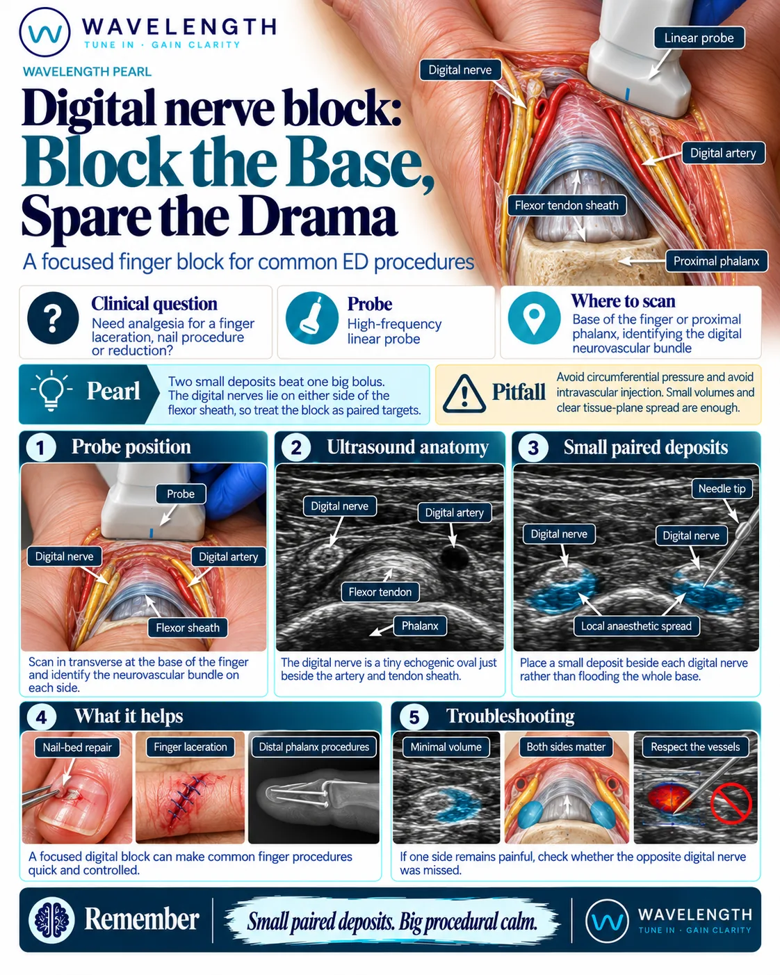
Scan images in the graphic are illustrations, not patient scans.
Before you block
- Record sensation on both sides of the finger and the tendon function before the block. A digital nerve injury must be documented first.
- Record consent. One to two millilitres on each side is usually enough.
- Lidocaine with adrenaline is safe in the fingers of healthy patients. A large multicentre study found no finger loss. Follow your trust's guidance, and avoid adrenaline where the circulation to the finger is already poor.
The scan
- Rest the hand palm up. Use plenty of gel, or a gel pad, over the base of the finger.
- Place a high-frequency linear probe transversely over the palmar surface of the proximal phalanx.
- Identify the bone, the bright flexor tendon in its sheath, and on each side the small digital artery with the nerve beside it. Colour Doppler finds the arteries.
- Insert the needle from the side of the finger, in plane or out of plane, and place a small deposit beside the nerve on one side.
- Repeat on the other side.
Reading the view
- Proximal phalanx. A curved bright line with a shadow below.
- Flexor tendon. A bright, fibrillar oval on the bone.
- Digital artery. A small black circle on each side, with flow on Doppler.
- Digital nerve. A tiny bright oval beside each artery.
- Correct spread. A small pool of fluid around each nerve.