A fractured neck of femur hurts with every movement, and older patients tolerate opioids poorly. The fascia iliaca block places a larger volume of local anaesthetic under fascia iliaca, away from the nerves, and relies on spread along the plane to reach the femoral nerve and, less reliably, the lateral femoral cutaneous nerve. UK hip fracture guidance supports a nerve block on arrival, and a Cochrane review found nerve blocks reduce pain on movement and the risk of delirium and chest infection.

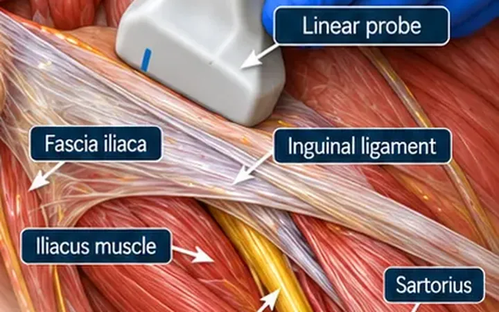
Scan images in the graphic are illustrations, not patient scans.
Before you block
- Record the neurovascular examination and look for other injuries.
- Record consent. Many patients with a hip fracture have delirium or dementia. Follow your trust's process for a patient who lacks capacity.
- Calculate the maximum safe dose for the patient's weight. This block uses 30 to 40 mL, so the dose decides the concentration.
- Attach monitoring and know where the lipid emulsion is.
The scan
- Lie the patient supine.
- Place the linear probe transversely just below the inguinal ligament and find the femoral artery, then the femoral nerve lateral to it.
- Slide lateral until you see the bright line of fascia iliaca lying on iliacus, about a third of the way from the artery to the anterior superior iliac spine.
- Insert the needle in plane from lateral to medial, aiming for the plane under fascia iliaca, well away from the nerve.
- Inject 2 to 3 mL and watch the fluid lift the fascia off iliacus. Then inject the rest, watching the fluid spread medially towards the nerve and laterally.
Reading the view
- Fascia iliaca. A bright line on top of iliacus.
- Iliacus. Dark, striated muscle under the fascia.
- Correct spread. A dark lens of fluid separating fascia iliaca from iliacus and spreading along it.
- Wrong layer. Fluid above the fascia, in the fat or around sartorius, does not reach the nerves.