A firm, thick Achilles tendon can be a chronic tendinopathy, a partial or complete tear, or a xanthoma, a deposit of cholesterol within the tendon. Achilles xanthomas are a recognised sign of familial hypercholesterolaemia, an inherited condition which causes early heart disease and is easily treated once found. The scan settles whether the fibres are continuous. That decides the next step.
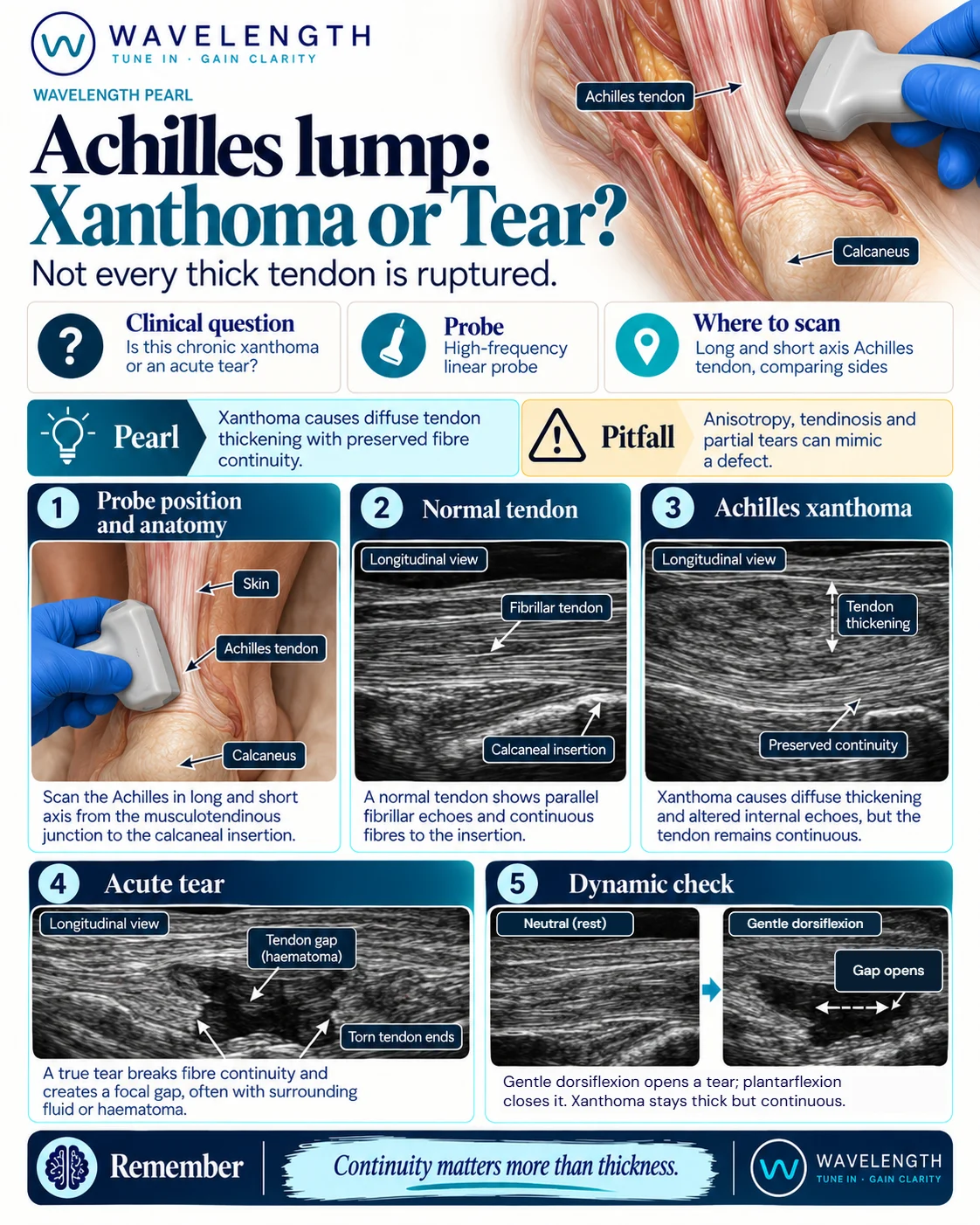
Scan images in the graphic are illustrations, not patient scans.
The scan
- Lay the patient prone with the feet over the end of the bed.
- Use a high-frequency linear probe with plenty of gel. Keep the beam at right angles to the tendon to avoid anisotropy.
- Scan in long axis from the muscle junction down to the insertion on the calcaneus.
- Scan in short axis along the same length and measure the front-to-back thickness at the thickest point.
- Scan the other side. Xanthomas are usually on both sides.
- If you suspect a tear, gently and passively dorsiflex the foot while watching in long axis.
Reading the view
- Normal: parallel bright fibres, continuous to the insertion. A normal Achilles is usually less than about 6 mm thick from front to back.
- Xanthoma: smooth, diffuse or spindle-shaped thickening with a darker, speckled texture, but the fibres stay continuous. Often on both sides, often painless.
- Tendinopathy: thickening, usually a few centimetres above the insertion, often painful, sometimes with new vessels on power Doppler.
- Complete tear: the fibres stop, with a gap filled by fluid or haematoma. The torn ends move apart with dorsiflexion and come together with plantarflexion. See the Achilles rupture pearl.
Action
A tear, or a patient who cannot plantarflex against resistance, follows your local Achilles rupture pathway. Thick but continuous tendons on both sides without an injury should prompt a fasting lipid profile and assessment for familial hypercholesterolaemia, through the patient's GP or a lipid clinic.