Pectoralis major tears happen mostly in young men lifting heavy weights, especially in the lowering phase of a bench press. Patients feel a pop, then pain, weakness and bruising over the front of the arm. The tear is often missed as a shoulder sprain. The tendon is superficial, so a linear probe can follow it to the humerus.
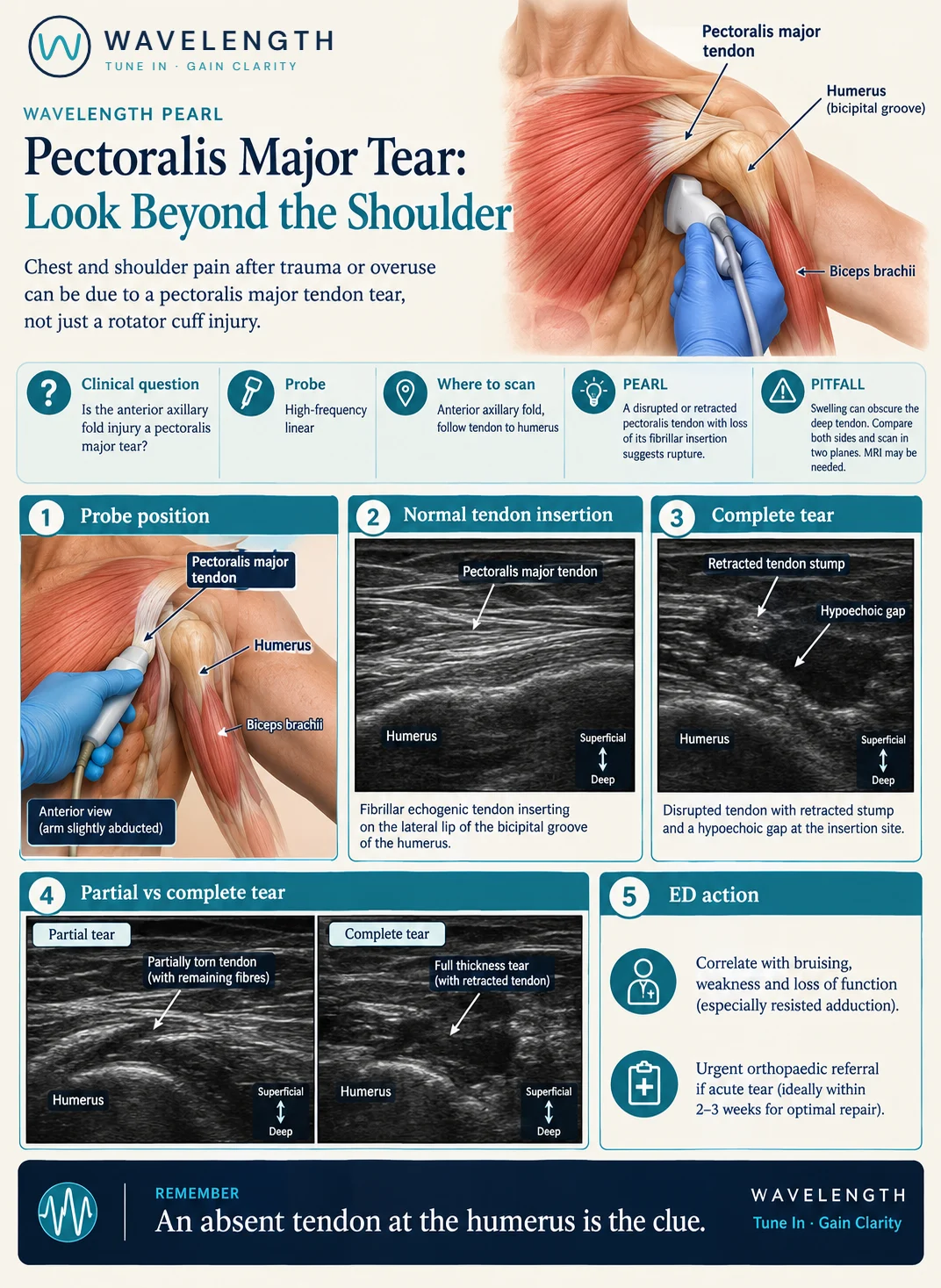
Scan images in the graphic are illustrations, not patient scans.
The scan
- Sit the patient up with the arm slightly away from the body and turned out.
- Use a high-frequency linear probe along the anterior axillary fold, following the muscle towards the arm.
- Follow the tendon to its insertion on the lateral lip of the bicipital groove of the humerus.
- Scan in long and short axis, and compare with the other side.
- Ask the patient to press the hand gently inwards (adduction) while you watch the tendon.
Reading the view
- Normal: a bright, fibrillar tendon running onto the humerus, moving as one with contraction.
- Complete tear: no tendon at the humerus, a dark gap filled with fluid or haematoma, and a retracted stump further towards the chest.
- Partial tear: some continuous fibres with a dark defect, or a tear at the muscle-tendon junction.
Action
Look for bruising, a thin or absent anterior axillary fold, and weakness of resisted adduction and internal rotation. Suspected complete tears need urgent orthopaedic referral, because repair in the first few weeks gives the best results.