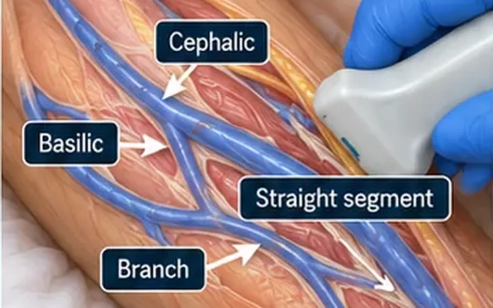
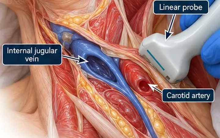
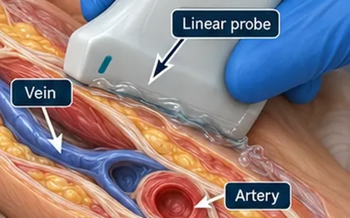
At a glance, a round black circle on the screen is a round black circle. A vein and the artery beside it look alike in short axis, especially in a shocked patient with a weak pulse. Cannulating the artery by mistake leads to bleeding, a painful haematoma and, if a drug goes in, possible harm to the hand. The bedside test takes two seconds: press.
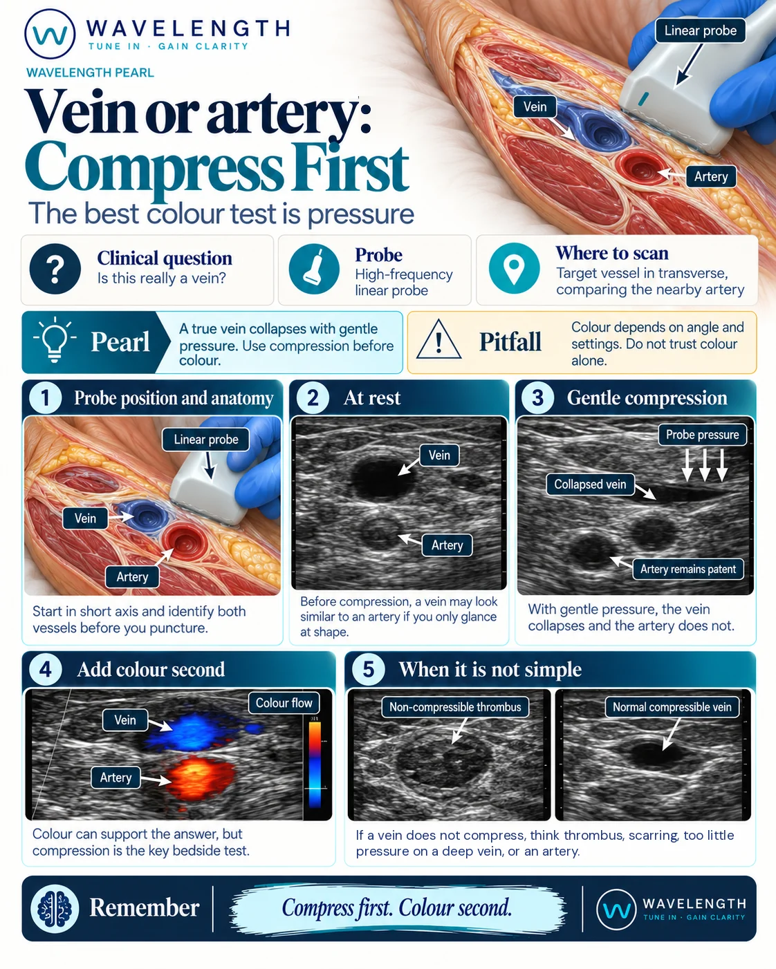
Scan images in the graphic are illustrations, not patient scans.
The scan
- Scan in short axis with a high-frequency linear probe. Find the target and the vessel beside it before you puncture.
- Look at both at rest. Shape alone does not separate them.
- Apply gentle, even pressure with the probe. The vein flattens and disappears. The artery stays round and pulsates.
- Release the pressure and let the vein refill before you puncture, using a light hand.
- Add colour Doppler second, if you need more. It supports the answer, but compression is the key bedside test.
Reading the view
- Vein. Collapses with gentle pressure. Thin wall, no pulsation.
- Artery. Stays open under the same pressure. Thicker wall, pulsates.
- Colour. Red and blue show the direction of flow relative to the probe, not the type of vessel. Change the angle or the settings and the colours change. Spectral Doppler gives more: arterial flow is pulsatile, venous flow is phasic or steady.
When the vein does not compress
If a vein does not compress, think of:
- Thrombus. Echogenic material in the lumen. Choose another vein.
- Scarring from past cannulation, with a thick, stiff wall.
- Too little pressure on a deep vein. Press a little more, then check again.
- An artery. Look for pulsation and check with Doppler.
Do not cannulate a vessel you cannot prove is a vein.