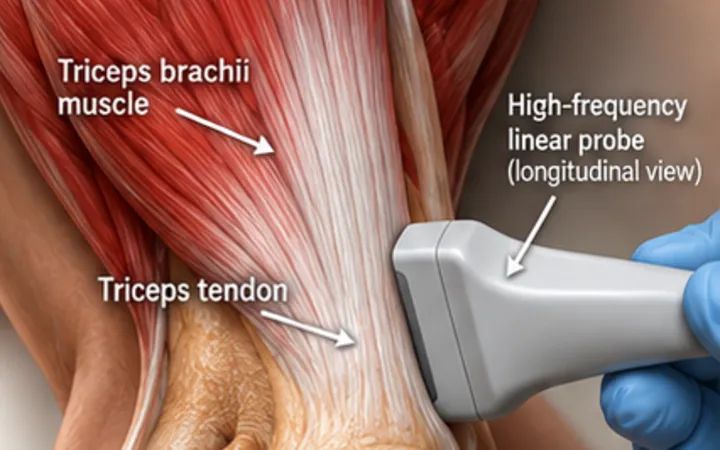
The extensor mechanism of the knee runs from the quadriceps tendon, through the patella and the patellar tendon, to the tibial tubercle. A rupture anywhere stops the patient straightening the knee. Swelling can hide the gap from your fingers, but not from the probe.
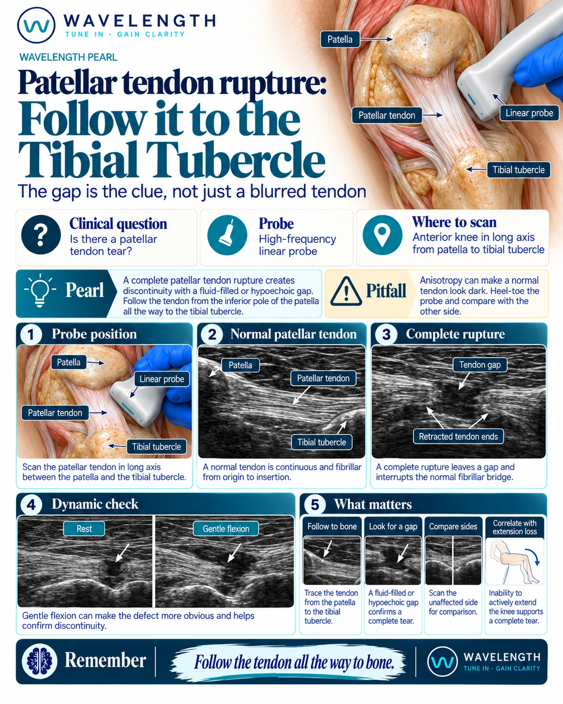
Scan images in the graphic are illustrations, not patient scans.
The scan
- Lie the patient supine with the knee slightly bent over a pillow.
- Place a high-frequency linear probe in long axis below the patella.
- Follow the tendon from the lower pole of the patella all the way to the tibial tubercle.
- Turn to transverse at any abnormal spot.
- If it is comfortable, bend the knee a little. A gap opens; an intact tendon stays continuous.
- Compare with the other knee.
Reading the view
- Normal: a bright, fibrillar, continuous tendon from patella to tubercle.
- Complete rupture: the fibres stop, with a dark gap of fluid or haematoma and wavy, retracted ends. The patella may sit higher than on the other side.
- Partial tear or tendinopathy: a thickened, darker tendon with some continuous fibres.
Action
A patient who cannot straight-leg raise, with a gap on ultrasound, needs urgent orthopaedic review: complete ruptures are usually repaired early. X-ray the knee to look for a high patella and a bony avulsion.