Wounds and burns over the back of the thumb, the first web space and the radial side of the back of the hand sit in the territory of the superficial branch of the radial nerve. It is a small, purely sensory nerve. In the forearm it runs beside the radial artery, under brachioradialis. A few millilitres around it, often combined with median and ulnar blocks, completes analgesia for the hand.
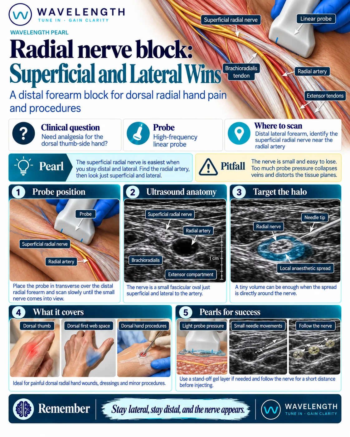
Scan images in the graphic are illustrations, not patient scans.
Before you block
- Record motor and sensory function of the hand before the block.
- Record consent and calculate the dose. Two to four millilitres is usually enough.
The scan
- Rest the forearm with the thumb side up.
- Place the linear probe transversely on the radial side of the forearm, in its middle third.
- Find the radial artery with colour Doppler, then look on its radial side for a small, bright, honeycomb oval under brachioradialis.
- Follow the nerve up and down to confirm it. Near the wrist it leaves the artery, passes under the brachioradialis tendon and becomes superficial, branching in the skin.
- Insert the needle in plane, keeping clear of the artery and the cephalic vein, and inject small amounts around the nerve.
Reading the view
- Radial artery. Round and pulsating, with colour on Doppler.
- Superficial radial nerve. A small honeycomb oval on the radial side of the artery.
- Brachioradialis. The muscle and tendon over the nerve.
- Correct spread. A small ring of fluid around the nerve.