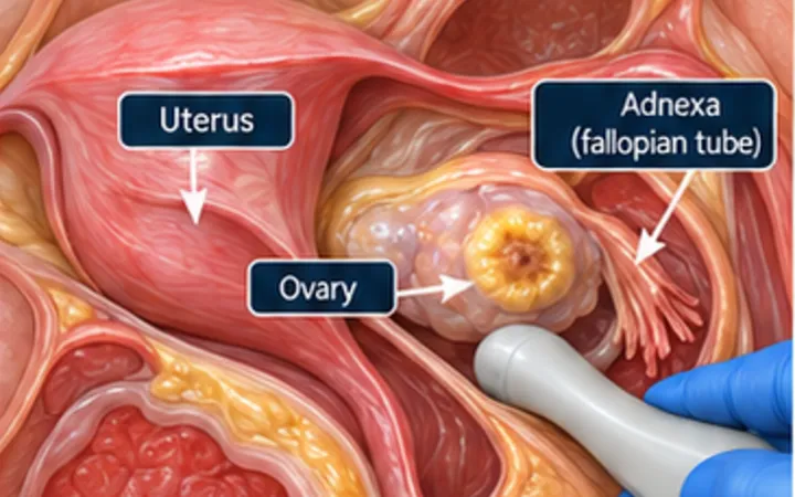
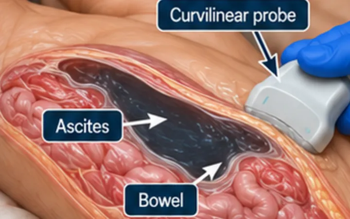
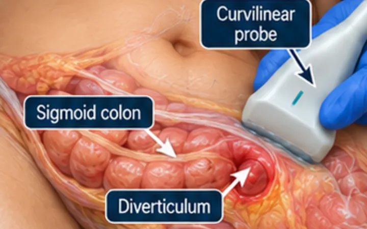
Pyelonephritis is a clinical and laboratory diagnosis. In most patients the kidney looks normal on ultrasound, and the subtle changes of infection are easy to miss at the bedside. The value of the scan lies elsewhere: an infected kidney behind a blockage needs urgent drainage, and that you can see.
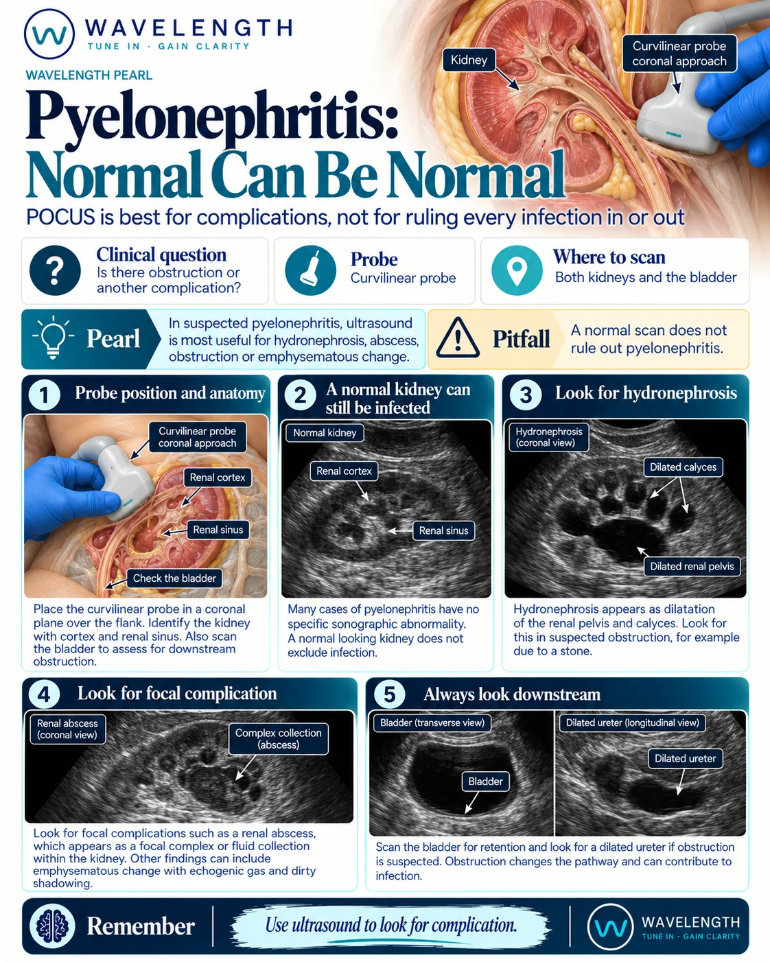
Scan images in the graphic are illustrations, not patient scans.
The scan
- Use the curvilinear probe in the flank, in the coronal plane, with the patient supine or on their side.
- Sweep each kidney fully in long and short axis. Compare the painful side with the other side.
- Look at the collecting system for dilatation.
- Look at the cortex for a focal collection.
- Scan the bladder for retention, and look behind it for a dilated ureter.
Reading the view
- Normal kidney: common in pyelonephritis. It does not exclude infection.
- Hydronephrosis: a dilated pelvis and calyces, black that branches. With fever, think of an obstructed, infected kidney. See the hydronephrosis pearl.
- Pyonephrosis: hydronephrosis with debris or layering echoes in the collecting system. See the pyonephrosis pearl.
- Abscess: a focal, rounded collection in or around the kidney, often with mixed echoes.
- Gas: bright echoes with dirty shadowing in the kidney suggest emphysematous pyelonephritis, mostly in people with diabetes. CT confirms it.
- Retention: a full bladder after voiding can cause obstruction and infection upstream.
Action
Hydronephrosis in a febrile patient, pyonephrosis, gas or an abscess needs senior and urology review the same day; an obstructed, infected kidney needs urgent drainage. Arrange formal imaging, usually CT, for these findings, for sepsis, for a single kidney, or when the patient is not improving after about 72 hours of treatment.