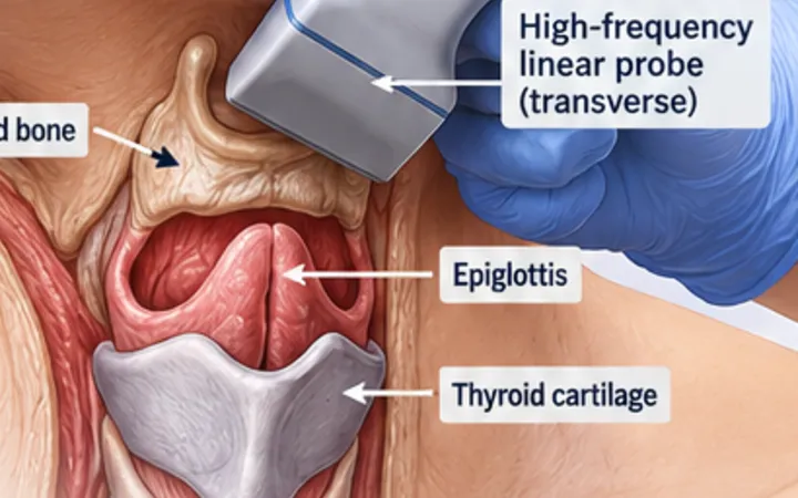
When a difficult airway is expected and you cannot feel the front of the neck, a minute of ultrasound can find the cricothyroid membrane. In obese patients and distorted necks, palpation often misses it, and ultrasound finds it much more often. The point is preparation: find it while oxygenation is stable, mark the skin, and have the rescue plan ready.
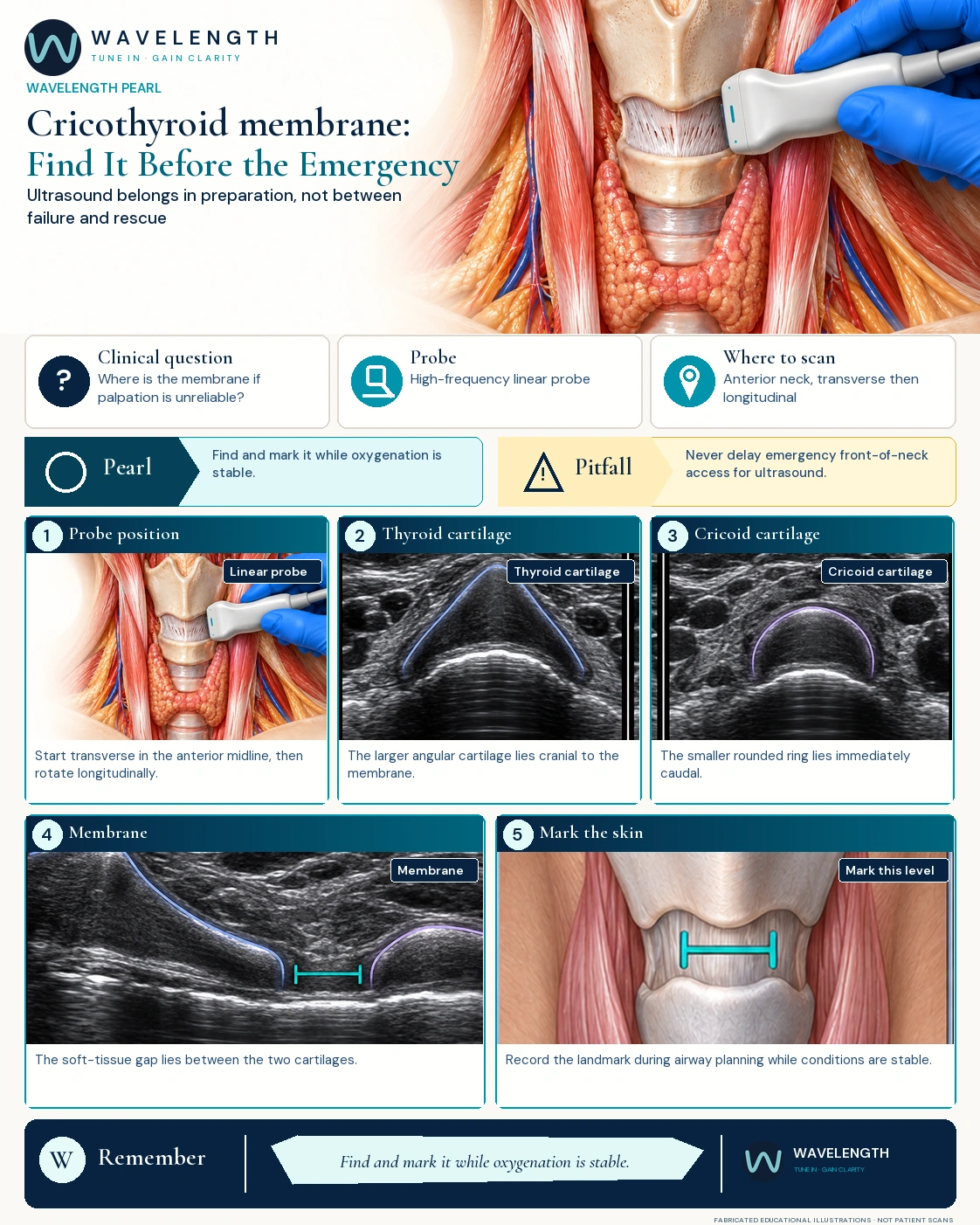
Scan images in the graphic are fabricated educational illustrations, not patient scans.
The scan
- Position the neck as your airway plan allows. Use a high-frequency linear probe, plenty of gel and a light hand.
- In transverse, find the thyroid cartilage: a larger, angular, dark structure with a bright line deep to it where air meets the airway lining.
- Slide down to the cricoid: a smaller, rounded dark ring.
- Turn the probe into the midline long axis. The membrane is the soft-tissue gap between the bottom of the thyroid cartilage and the top of the cricoid.
- Mark the centre of the membrane, and the midline, on the skin with the neck in the position you will use for the procedure.
- Check the mark again if the head or neck moves a lot.
Use it safely
- It helps most when the landmarks are hard to feel: obesity, neck swelling, previous surgery or radiotherapy.
- Ultrasound does not replace the difficult airway plan, the equipment, a trained assistant or the scalpel technique.
- In a can't intubate, can't oxygenate emergency, do not delay front-of-neck access to fetch or use a scanner.
Action
If the landmarks are difficult and time allows, find and mark the membrane during airway preparation, and tell the team the mark may need checking if the patient is moved.