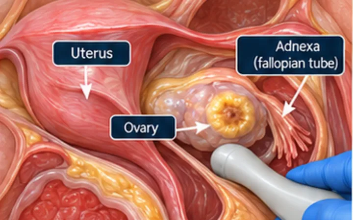
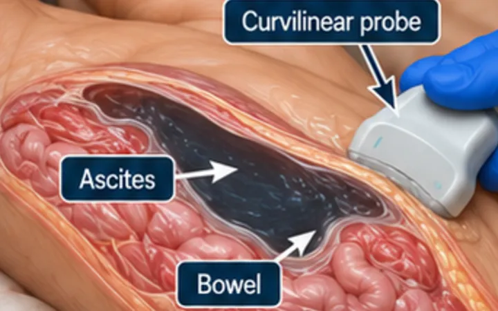
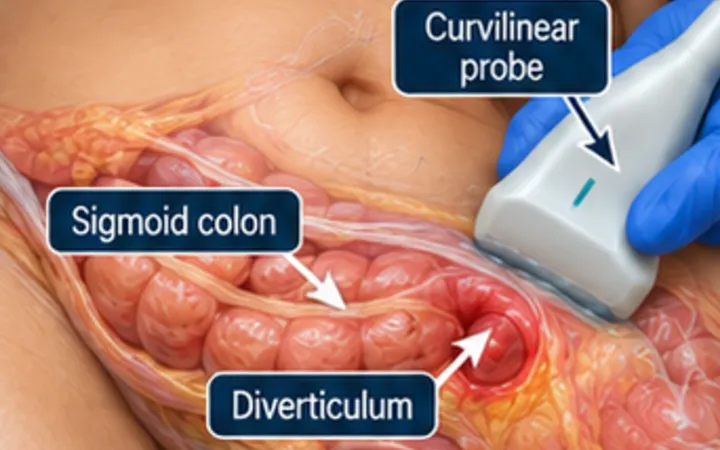
Ultrasound is not the definitive test for a perforation, but it can show free air at the bedside. Seeing gas is not enough, because bowel always contains gas. The clue is gas lying directly against the front of the peritoneum.
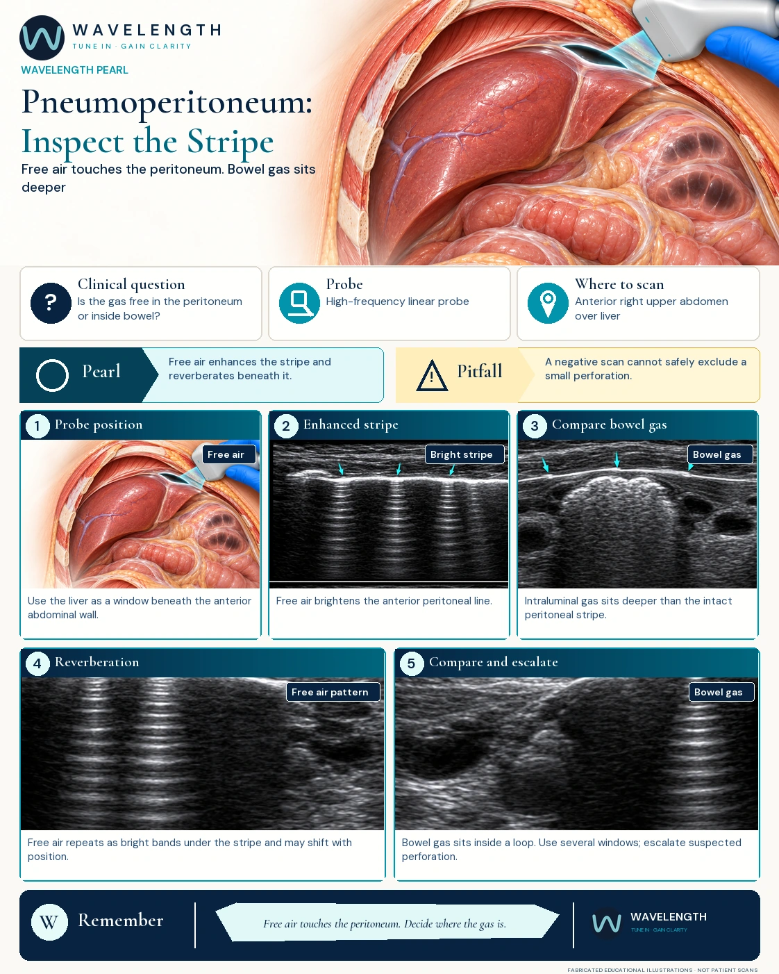
Scan images in the graphic are fabricated educational illustrations, not patient scans.
The scan
- Lie the patient flat. Place a high-frequency linear probe on the right upper abdomen, over the liver, which gives a clean window under the abdominal wall.
- Find the normal peritoneal stripe: a thin bright line between the abdominal wall and the liver.
- Look for a stripe which is brighter and thicker than usual, with reverberation lines or a dirty shadow beneath it.
- Change the angle and, if it is safe, the patient's position. Free air can move; gas inside bowel stays with the bowel.
- Look for support: free fluid, bright bubbles in fluid, sluggish bowel or local inflammation.
Reading the view
- Free air: the peritoneal stripe is abnormally bright, and repeats beneath itself as reverberation, because gas touches the peritoneum.
- Bowel gas: the thin peritoneal line stays visible on top, with the gas artefact deeper, inside a loop.
- A normal scan does not rule out a perforation, especially with a small amount of air or a poor window.
Action
If you suspect free air in an unwell patient, escalate urgently and follow your surgical and CT pathway. Do not let a normal bedside scan overrule strong clinical concern.