A varicocele is a cluster of dilated veins around the spermatic cord. It is common, usually painless or a dull ache, and usually on the left, because the left testicular vein drains at a right angle into the left renal vein. Most are not urgent. Ultrasound confirms the diagnosis and, as importantly, checks the testis and the side.
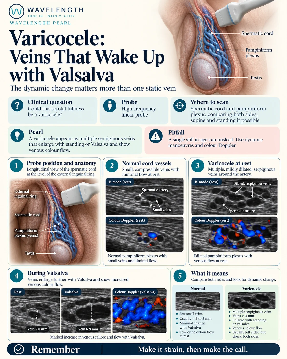
Scan images in the graphic are illustrations, not patient scans.
The scan
- Explain the scan, offer a chaperone and record the offer.
- With the patient lying flat, use a high-frequency linear probe above the testis, along the spermatic cord.
- Measure the widest vein at rest, and add colour Doppler with a low scale.
- Ask the patient to bear down (Valsalva), and watch the veins and the colour. Repeat standing if you can.
- Scan both sides, and both testes.
Reading the view
- Normal: a few small veins, usually under about 2 to 3 mm, with little change on straining.
- Varicocele: several tortuous veins over about 3 mm, which enlarge and fill with reversed flow on Valsalva or standing.
- Testes: compare their size. A smaller testis on the varicocele side matters for fertility referral.
Action
Most varicoceles are referred routinely to urology or the GP, especially with pain, a smaller testis or fertility concerns. A new varicocele on the right, one on both sides that appeared suddenly, or one that does not empty when the patient lies down, can come from a mass pressing on the veins higher up: arrange imaging of the abdomen and kidneys.