A painful lump in the neck is a common emergency presentation, in children and adults. The question is whether it is an inflamed lymph node, which settles with treatment, or a collection, which may need drainage. A linear probe answers that quickly for superficial lumps. The same scan can raise the possibility of something more serious.

Scan images in the graphic are illustrations, not patient scans.
The scan
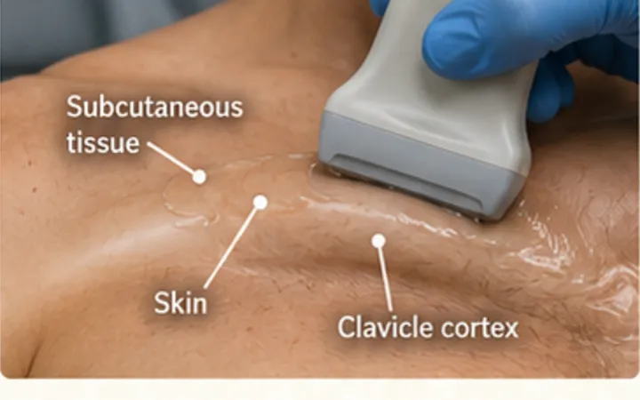
- Use a high-frequency linear probe. Sit the patient up with the neck turned slightly away.
- Find the lump and the landmarks around it: the sternocleidomastoid muscle, the carotid artery and the internal jugular vein.
- Scan the lump in long and short axis. Measure it in three directions.
- Look for a central bright hilum, the shape, and the texture inside.
- Add colour Doppler: a node usually has flow entering through the hilum.
- Press gently. Pus often swirls.
Reading the view
- Reactive node: oval, smooth, with a dark rim of cortex around a bright central hilum, and flow entering at the hilum.
- Abscess: round or irregular, with no hilum, complex fluid with debris, posterior enhancement and no flow in the centre. The contents may swirl with pressure.
- Suppurative node: a node starting to liquefy, with a dark centre and a lost hilum. Common in children.
- Worrying node: round, with no hilum, or with a dark or cystic centre in an adult. Think of a metastatic or tuberculous node.
Action
Stridor, trismus, drooling or difficulty swallowing need urgent senior and ENT review, whatever the scan shows. A collection may need drainage by ENT or under ultrasound guidance. A persistent unexplained neck lump, especially in an adult, needs referral on your local urgent suspected cancer pathway. See the abscess pearl for the swirl sign.