A swollen, red point of the elbow could be a bursitis, cellulitis, an abscess or a joint effusion. They sit in different layers, and the layer changes what you do next. The olecranon bursa lies just under the skin, on top of the olecranon and the triceps insertion. The elbow joint lies deeper.

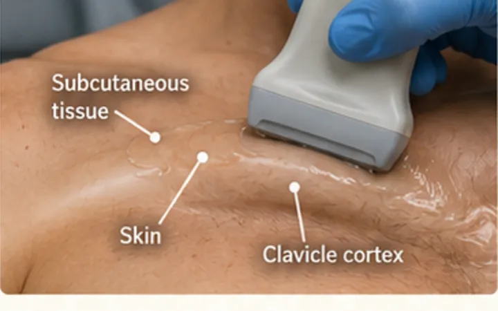
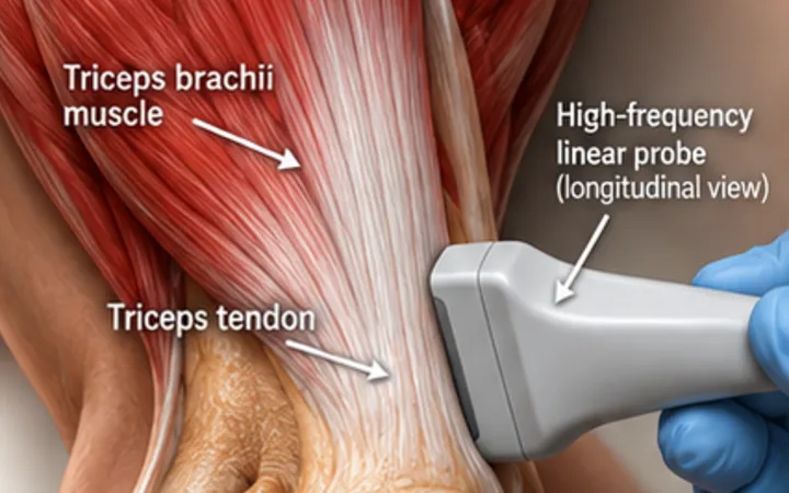
Scan images in the graphic are illustrations, not patient scans.
The scan
- Rest the elbow bent on a pillow. Use a high-frequency linear probe with plenty of gel and almost no pressure, or the bursa empties under the probe.
- Scan over the point of the elbow in long axis and transverse.
- Name the layers from the top: skin and fat, the bursa, the triceps tendon and the bright cortex of the olecranon.
- Decide where any fluid is, then what it looks like.
- Look deeper, at the posterior recess of the joint, to make sure the joint itself is not full of fluid.
Reading the view
- Normal: the bursa is collapsed, with little or no visible fluid.
- Simple bursitis: clear, dark fluid in the bursa, above the triceps tendon.
- Complex bursitis: debris, septations or a thick wall, often with increased blood flow around it on power Doppler. Infection is more likely, but this appearance is not proof.
- Cellulitis: swollen, cobblestoned fat without a collection in the bursa.
Action
If infection is possible, aspirate under ultrasound guidance and send the fluid for microscopy, culture and crystals. If the fluid is in the joint, not the bursa, treat it as a possible septic arthritis.