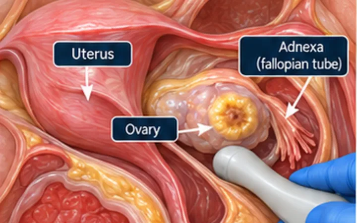
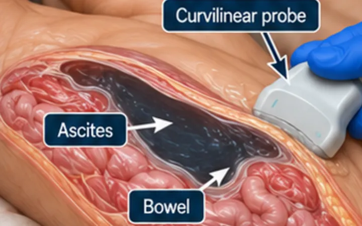
Free fluid in the pelvis does not promise to sit in the middle of one suprapubic image. The pelvic view of the eFAST has to sweep through the dependent spaces behind and beside the bladder, in both sagittal and transverse.
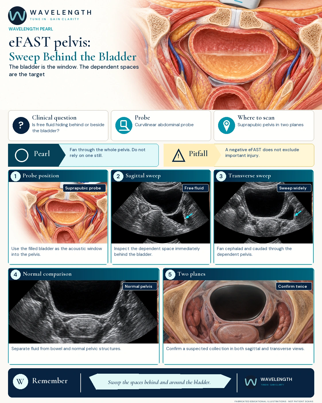
Scan images in the graphic are fabricated educational illustrations, not patient scans.
The scan
- Place the curvilinear probe just above the pubic symphysis and angle down into the pelvis.
- In sagittal, find the bladder and sweep from one side, through the midline, to the other. Look behind the bladder and, in women, around and behind the uterus.
- Turn 90 degrees into transverse. Fan up and down through the whole bladder and the space behind it.
- Set the depth so the space behind the bladder is fully on the screen.
- Save clips of the sweeps, not one still image.
Reading the view
- Free fluid is usually black or dark grey, and collects behind the bladder in men (the rectovesical pouch) and behind the uterus in women (the pouch of Douglas). Fresh blood can be grey.
- In women, a small amount of fluid can be normal. Read the amount, the place, the mechanism and the observations together.
- An empty bladder gives a poor window. A poor pelvic view does not make the whole scan negative.
Action
In unstable trauma, read the pelvic view with the rest of the eFAST and your trauma pathway. Repeat it if the observations change. A negative scan must not delay definitive care when suspicion stays high.