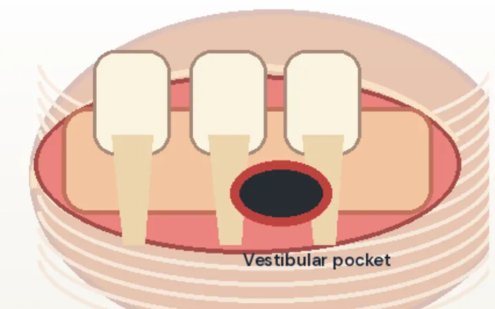
Pyomyositis is a bacterial infection of skeletal muscle, usually Staphylococcus aureus. It is commoner in the tropics, but in the UK it affects people with diabetes, immunosuppression, injecting drug use or recent muscle injury, and children. It starts as diffuse swelling and, over one to three weeks, forms an abscess inside the muscle. The skin can look almost normal, so it is easily called a strain, a DVT or cellulitis.
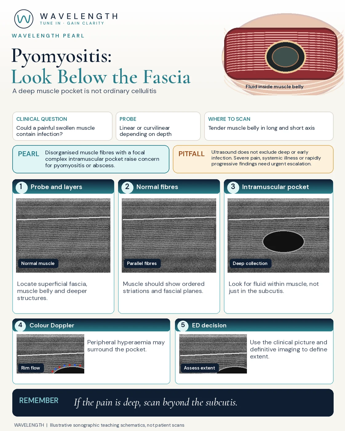
Scan images in the graphic are illustrations, not patient scans.
The scan
- Use a high-frequency linear probe for superficial muscles, and a curvilinear probe for the deep thigh or buttock.
- Find the skin, the fat, the bright fascia and the muscle below it.
- Scan the tender muscle in long and short axis, through its full depth.
- Press gently and add colour Doppler.
- Compare with the same muscle on the other side.
Reading the view
- Normal muscle: dark, with fine bright striations and bright fascial lines.
- Early infection: a swollen muscle that has lost its normal striations.
- Abscess: a hypoechoic or complex pocket inside the muscle, below the fascia, often with debris, swirl on pressure and flow in its rim.
- Not this: cobblestoning in the fat above the fascia is cellulitis, and fluid along the deep fascia raises concern for necrotising fasciitis.
Action
Take blood cultures and treat sepsis. MRI, or CT, shows the extent. A collection needs surgical or orthopaedic review for drainage, with antibiotics guided by the cultures.