A middle-aged player feels a sudden pop or a kick in the back of the calf while pushing off. The calf swells and hurts. This is "tennis leg", a tear where the medial head of gastrocnemius meets soleus. It looks like a DVT on the outside, and ultrasound tells them apart.
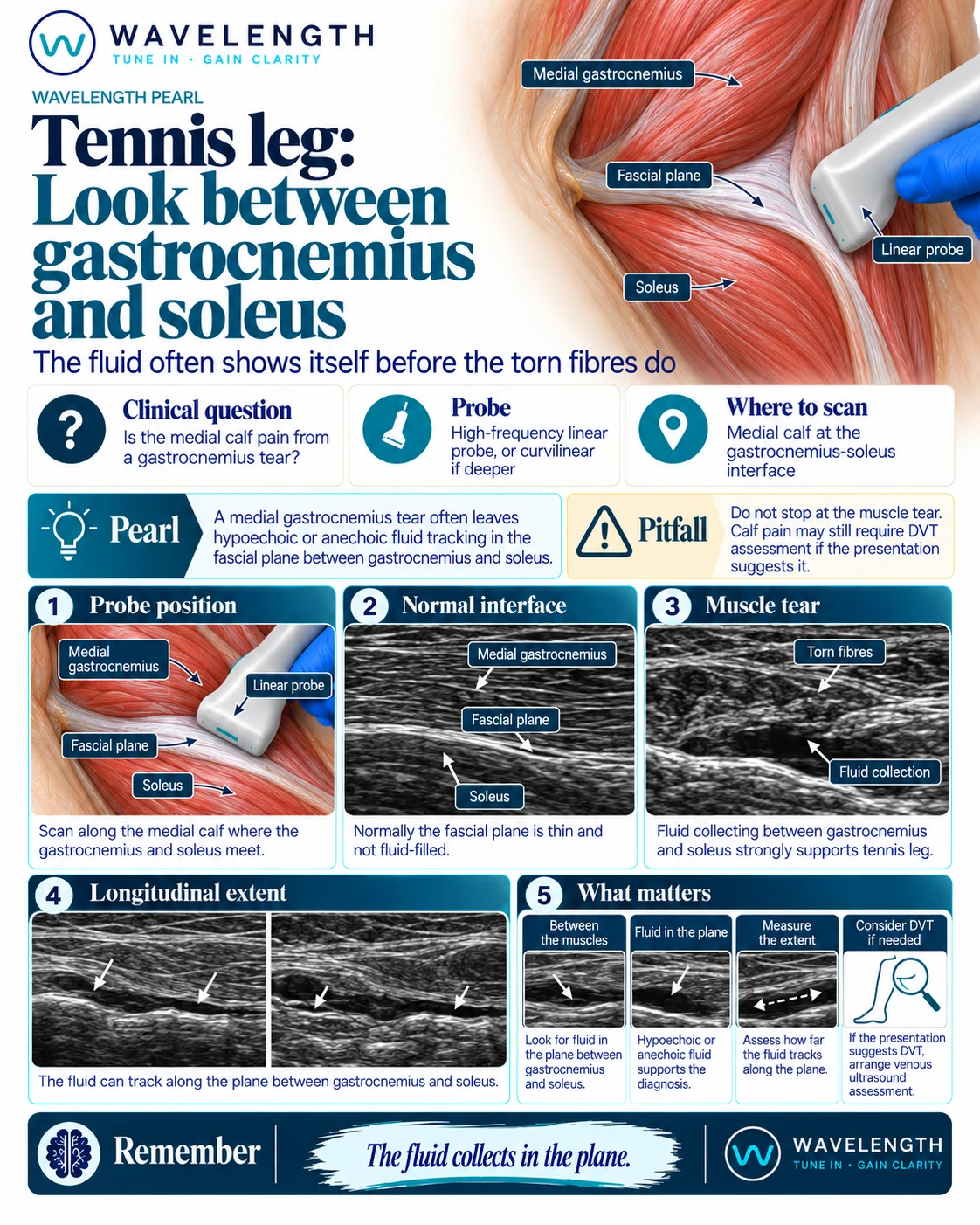
Scan images in the graphic are illustrations, not patient scans.
The scan
- Lie the patient prone, or on their side, with the calf relaxed.
- Place a high-frequency linear probe in long axis over the medial calf. Use the curvilinear probe for a large calf.
- Find the medial head of gastrocnemius, soleus beneath it, and the thin bright plane between them.
- Follow the tapering end of gastrocnemius down the calf, where tears usually happen.
- Look for torn fibres and dark fluid in the plane, then measure how far the fluid runs.
- Compare with the other calf.
Reading the view
- Normal: a thin, bright fascial plane between the two muscles, with no fluid.
- Tennis leg: disrupted fibres at the end of the medial gastrocnemius, with a dark collection tracking along the plane.
- Large collections can track a long way down the calf.
Action
Most tennis leg is managed with analgesia, early movement and physiotherapy. Assess for DVT when it is clinically indicated. Very large or expanding haematomas, or a tense calf, need senior review.