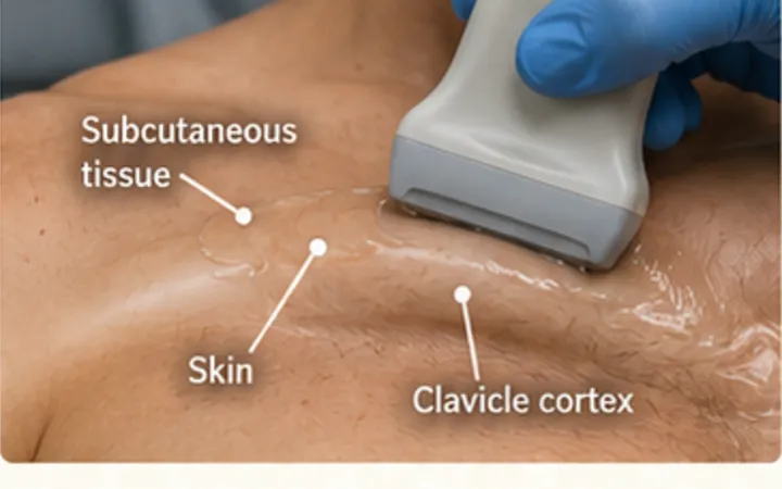
Chest X-rays miss many rib fractures, especially at the front, the sides and the costal cartilage. Ultrasound has found more fractures than X-ray in several studies, because you scan exactly where it hurts. The number of fractures, the patient's age and the lungs underneath decide the plan.
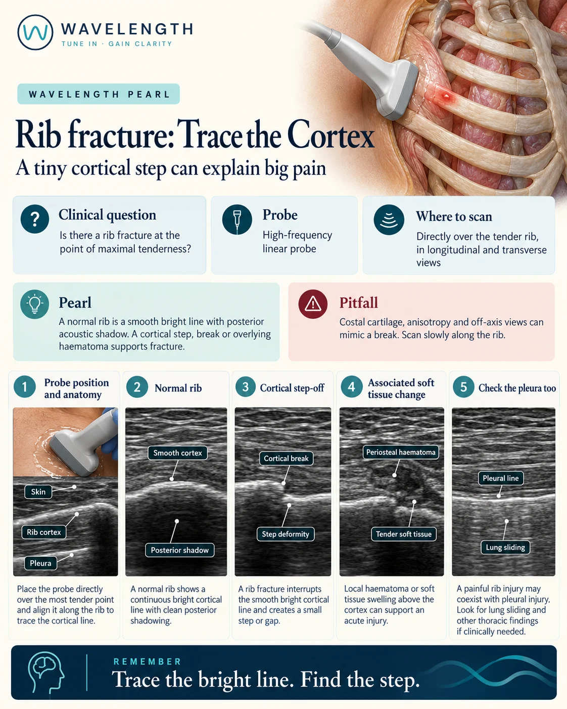
Scan images in the graphic are illustrations, not patient scans.
The scan
- Ask the patient to point to the most tender spot.
- Use a high-frequency linear probe along the long axis of the rib, so the cortex is a long bright line with a shadow beneath it.
- Slide slowly along the rib through the painful area, then turn to short axis over any abnormality.
- Compare with the rib above and below.
- Look under the rib, between the ribs, for the pleural line and lung sliding.
Reading the view
- Normal rib: a smooth, continuous bright line with a clean shadow beneath.
- Fracture: a step, a gap or a break in the bright line, at the point of tenderness.
- Supporting signs: a haematoma over the cortex, and pain as the probe passes over it.
- Costal cartilage: darker than bone, with no shadow. A break here is invisible on X-ray but can be seen on ultrasound.
- Underneath: absent lung sliding suggests a pneumothorax; fluid above the diaphragm suggests a haemothorax.
Action
Treat the patient, not the crack: analgesia that lets them breathe and cough. Older patients and anyone with several fractures, lung disease or anticoagulation need your local chest injury pathway, often with a regional block and admission. A pneumothorax or haemothorax changes management at once.