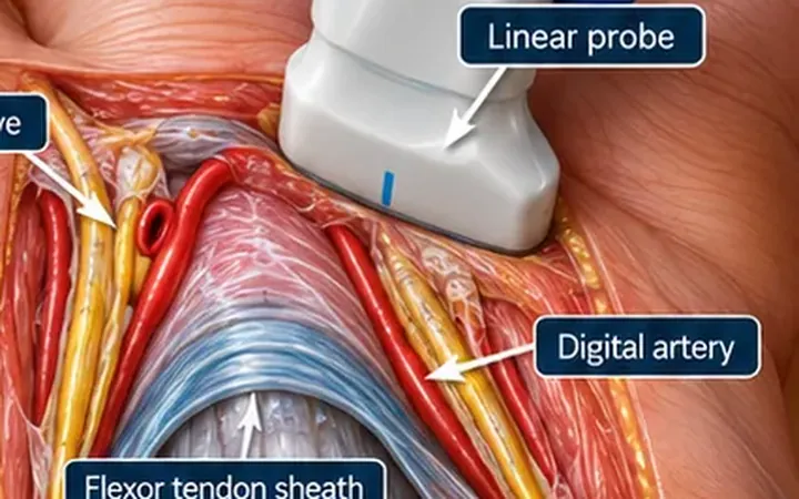
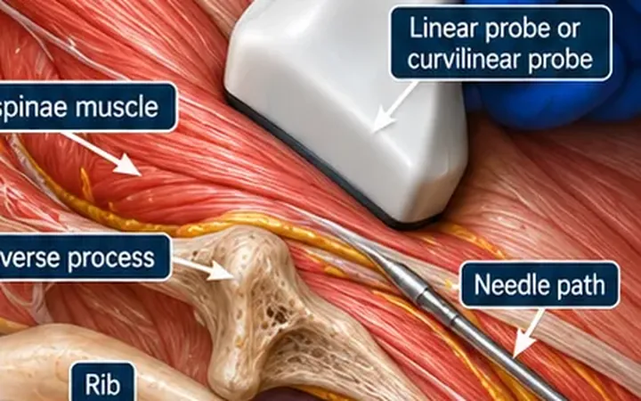
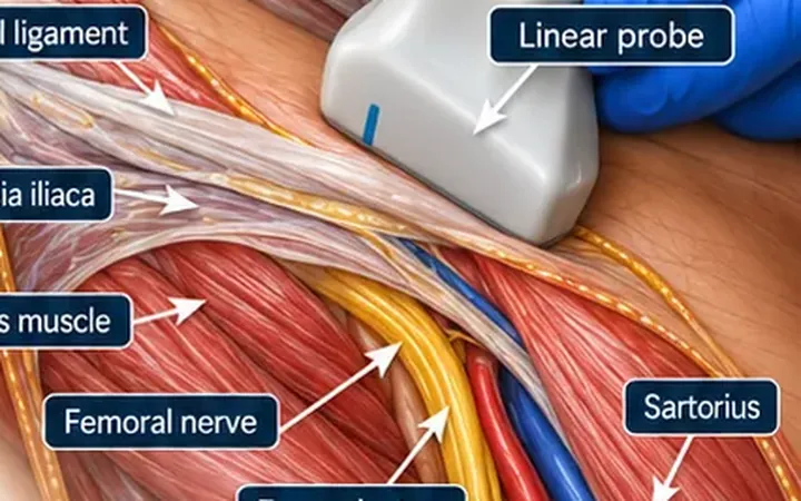
The front of the abdominal wall around the midline receives its supply from the anterior branches of the lower thoracic nerves. These run between rectus abdominis and the posterior rectus sheath before turning forward through the muscle. A rectus sheath block places local anaesthetic in this space. It treats pain from the abdominal wall, not from inside the abdomen. Most of the evidence comes from operating theatres, for midline and umbilical incisions.
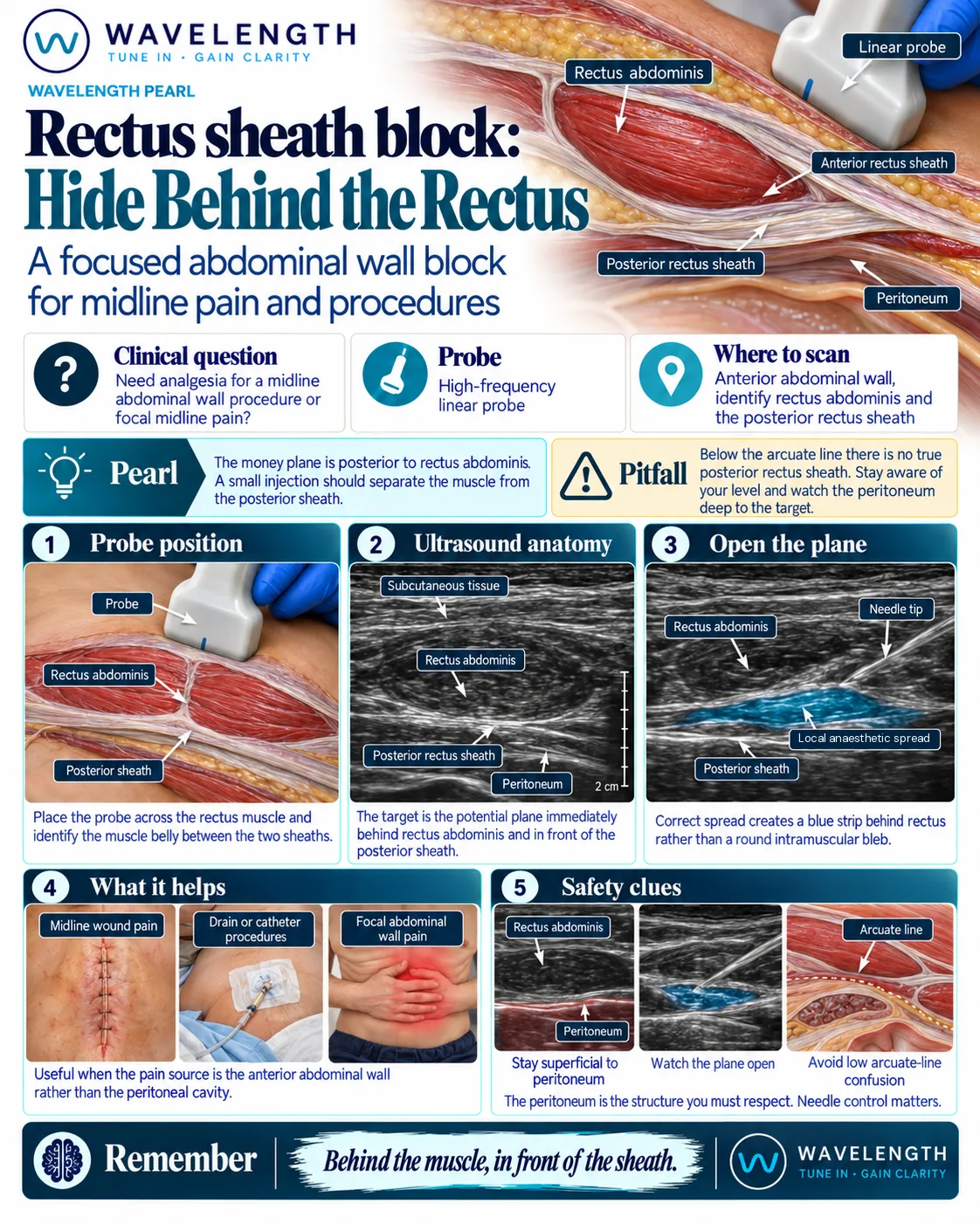
Scan images in the graphic are illustrations, not patient scans.
Before you block
- Be sure the pain comes from the abdominal wall. A block can hide the signs of a problem inside the abdomen.
- Record consent, calculate the maximum safe dose (a midline wound needs both sides), attach monitoring and know where the lipid emulsion is.
The scan
- Lie the patient supine.
- Place the linear probe transversely just lateral to the umbilicus, above the arcuate line.
- Identify rectus abdominis, an oval muscle between the bright anterior and posterior sheaths, and the peritoneum deep to the posterior sheath.
- Use colour Doppler to find the inferior epigastric vessels, which run on the back of the muscle.
- Insert the needle in plane from the lateral side, through the muscle, until the tip lies just above the posterior sheath.
- Inject 1 to 2 mL and watch the fluid separate the muscle from the posterior sheath. Then inject the rest. Repeat on the other side for a midline wound.
Reading the view
- Rectus abdominis. An oval muscle between two bright lines.
- Posterior rectus sheath. The deeper bright line. The peritoneum lies just beneath it.
- Correct spread. A dark strip of fluid between the back of the muscle and the posterior sheath.
- Wrong layer. A round swelling within the muscle.