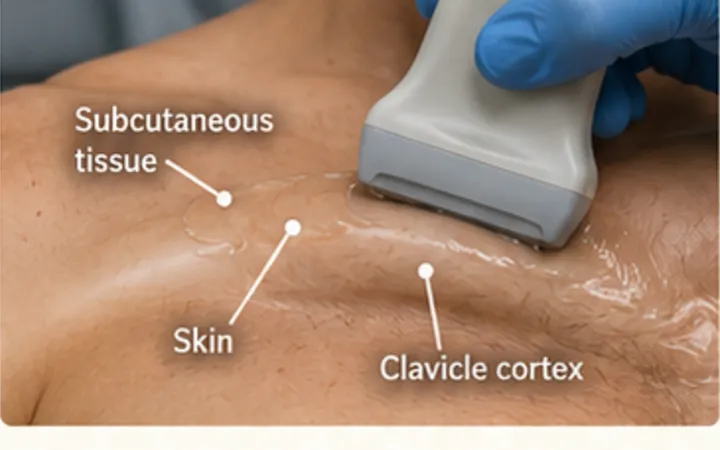
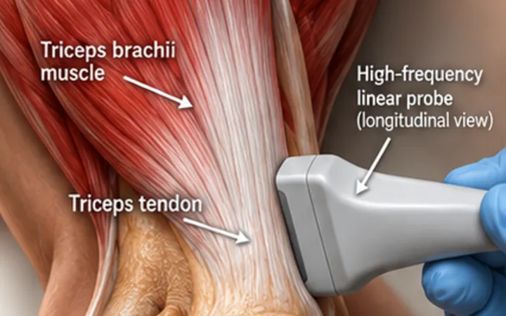
Ultrasound cannot see through adult bone, but it shows the surface very well. Instead of looking for a fracture line inside the bone, trace the bright cortex and look for a break in it. The distal radius in children is the best-studied example, but the same method works on any bone surface you can reach.
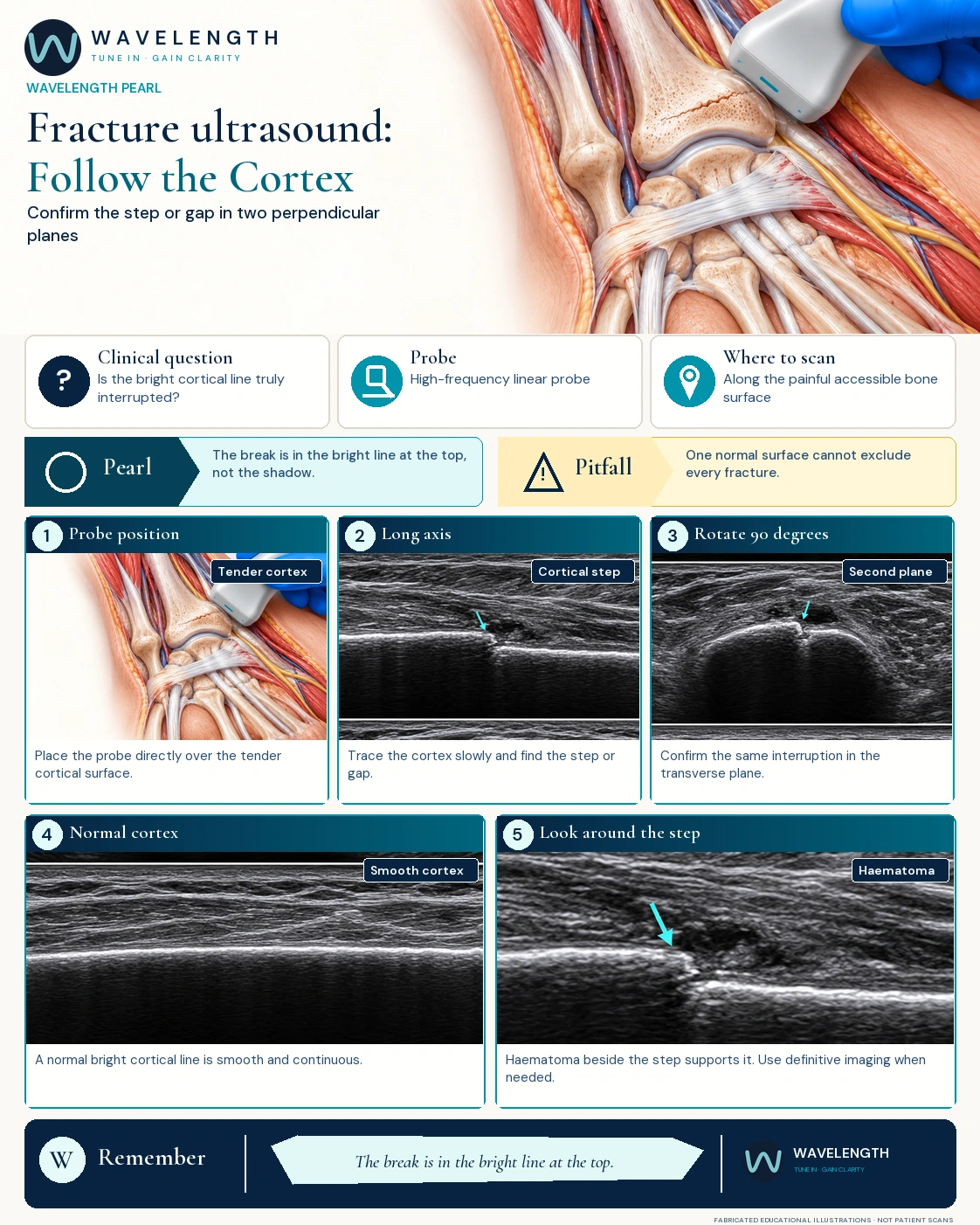
Scan images in the graphic are fabricated educational illustrations, not patient scans.
The scan
- Use a high-frequency linear probe and plenty of gel. Start away from the most painful spot.
- Find the bright cortex and slide slowly along it in long axis.
- At any step, gap, buckle or angle, turn the probe 90 degrees and find the same break in transverse.
- Look for haematoma beside it, a lifted periosteum and tenderness under the probe.
- Compare with the other side when the anatomy is complex.
Reading the surface
- Direct sign: a break, step or buckle in the cortex, seen in two planes.
- Supporting signs: haematoma, soft tissue swelling, a lifted periosteum and point tenderness.
- Mimics: growth plates, ossification centres and nutrient vessels in children, and old injuries in adults, can look like a break. Know the normal anatomy and compare sides.
Action
Use ultrasound when it answers a focused question at the bedside. Immobilise, and arrange X-ray or follow-up whenever clinical concern remains.