After a fall on the outstretched hand, a raised posterior fat pad on the lateral X-ray is the classic clue to a hidden elbow fracture. Ultrasound shows the same thing directly: fluid in the posterior recess, lifting the fat pad out of the olecranon fossa.
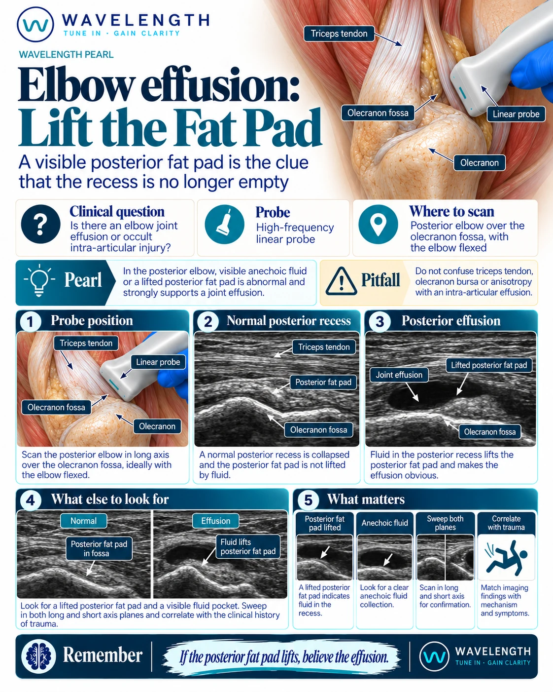
Scan images in the graphic are illustrations, not patient scans.
The scan
- Bend the elbow to about 90 degrees, with the forearm resting on the patient's lap or a pillow.
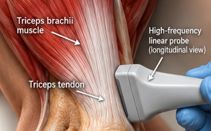
- Place a high-frequency linear probe in long axis over the back of the elbow, above the olecranon, along the line of the humerus.
- Find the triceps tendon, the posterior fat pad and the dip of the olecranon fossa.
- Look for dark fluid in the fossa, lifting the fat pad off the bone.
- Sweep in long axis and transverse, and compare with the other elbow.
Reading the view
- Normal: the fat pad sits down in the olecranon fossa, with no visible fluid.
- Effusion: dark fluid in the posterior recess lifts the fat pad. Fluid in the joint is abnormal.
- After trauma: an effusion strongly suggests an injury inside the joint, but the fracture line itself may not be visible on ultrasound.
Action
After trauma, an effusion means treating it as a possible occult fracture: immobilise, X-ray and arrange follow-up through your fracture pathway. Without trauma, a hot elbow with an effusion needs aspiration for possible septic arthritis.