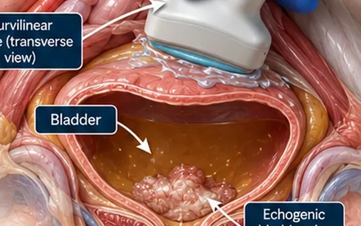
A patient who cannot pass urine, a confused older person with lower abdominal pain, or a catheter that seems not to drain: each raises one question, is the bladder full? A bedside scan answers it in a minute. The kidneys answer the next question, how far back the obstruction reaches.
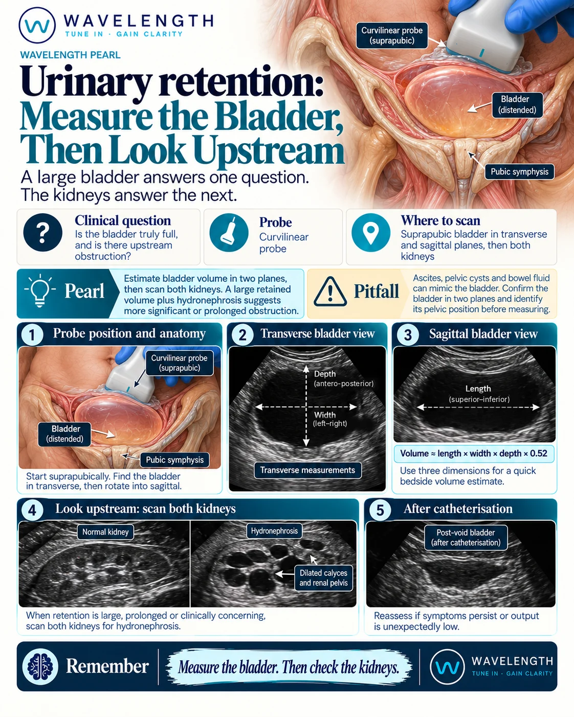
Scan images in the graphic are illustrations, not patient scans.
The scan
- Place the curvilinear probe just above the pubic symphysis, transverse, angled down into the pelvis.
- Find the widest transverse view. Measure the width, side to side, and the depth, front to back.
- Rotate to sagittal, marker to the head. Measure the length, head to foot.
- Estimate the volume: length × width × depth × 0.52.
- Scan both kidneys for hydronephrosis.
- After catheterisation, rescan the bladder to confirm it has emptied.
Reading the view
- Retention: a large, round, thin-walled bladder rising out of the pelvis. A volume of several hundred millilitres in a patient who cannot void supports the diagnosis. Record the number and compare it with the volume drained.
- Upstream effect: hydronephrosis on both sides with a large bladder suggests longstanding or high-pressure obstruction. Check the creatinine.
- Catheter in place but bladder full: the catheter is blocked or misplaced. Look for the balloon. A balloon in the urethra or outside the bladder needs urgent attention.
After the catheter
Expect a brisk diuresis after relief of chronic retention with renal impairment, and monitor urine output and electrolytes. If symptoms persist or output is lower than expected, rescan the bladder and the catheter balloon.