When a patient has gallstones and jaundice, abnormal liver tests or pancreatitis, the question is whether the common bile duct is dilated. The duct runs beside the portal vein and the hepatic artery, and the three look alike on greyscale. Colour Doppler sorts them in seconds.
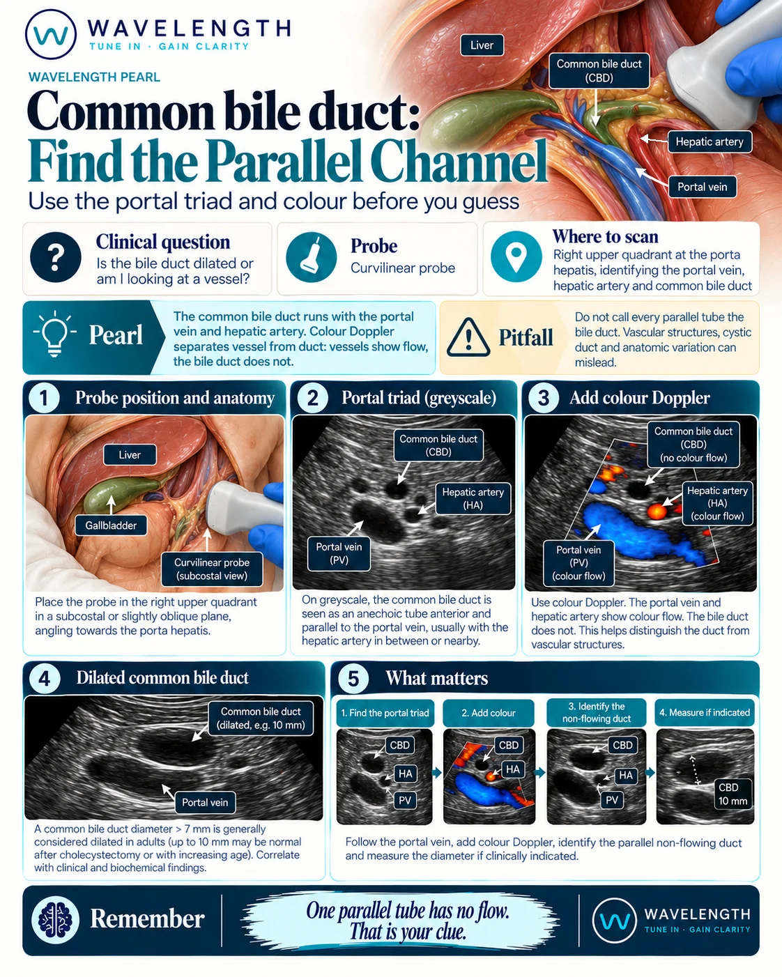
Scan images in the graphic are illustrations, not patient scans.
The scan
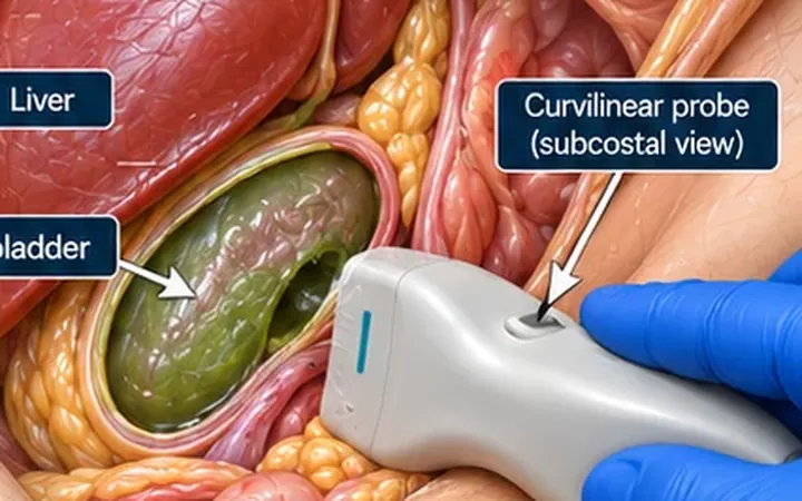
- Find the gallbladder with the curvilinear probe and follow its neck towards the porta hepatis.
- Find the portal vein, the large black vessel with bright walls entering the liver.
- Look just anterior to it. In cross-section the portal vein and the two smaller tubes in front of it form a face with two ears: the bile duct on the patient's right and the hepatic artery on the left.
- Rotate along the length of the duct so it runs across the screen, anterior and parallel to the portal vein.
- Add colour Doppler with a low velocity scale. The portal vein and hepatic artery fill with colour. The bile duct stays black.
- Measure the duct from inner wall to inner wall, at its widest point.
Reading the view
- Normal: most references treat a duct up to 6 mm as normal in adults, and some use 7 mm.
- Older patients: the duct widens with age. A common rule allows about 1 mm more for each decade over 60.
- After cholecystectomy: a duct up to 10 mm can be normal.
- Dilated: a wide duct with gallstones, jaundice or deranged liver tests suggests obstruction and needs further imaging.
What the scan does not tell you
POCUS rarely shows a stone in the distal duct, which hides behind the duodenum. A normal-calibre duct does not exclude a stone. Correlate with bilirubin, liver tests and the clinical picture, and request formal imaging when obstruction is suspected.