A patient with visible haematuria or clot retention often has echogenic material in the bladder. Clot, debris and a bladder tumour all look bright. Two quick checks, movement and colour, separate most of them at the bedside.
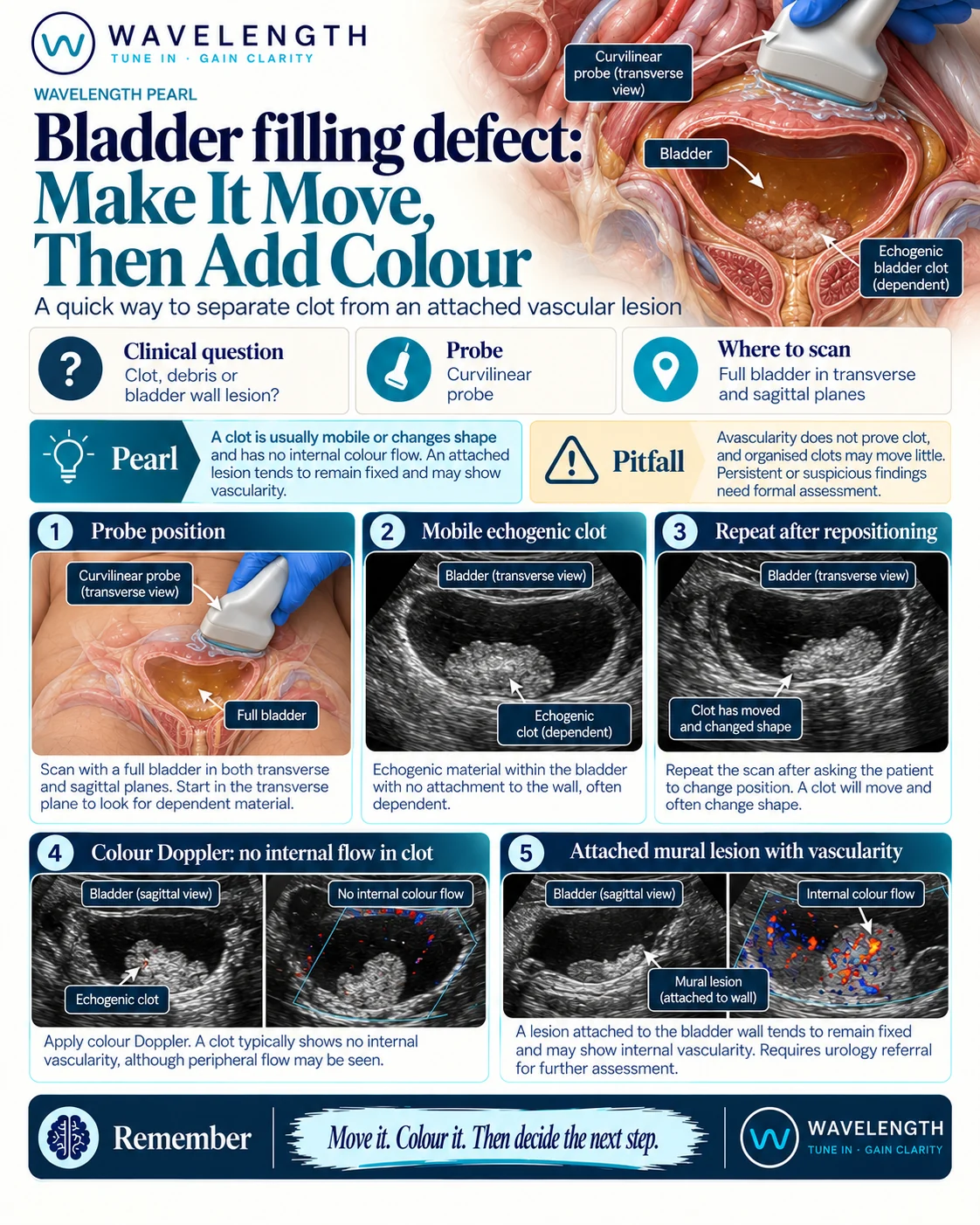
Scan images in the graphic are illustrations, not patient scans.
The scan
- Scan the bladder with the curvilinear probe just above the pubic symphysis, in transverse and then sagittal. The scan works best with some urine in the bladder.
- Find the echogenic material and note where it sits.
- Ask the patient to roll onto one side, or tilt the bed, and scan again. Watch whether the material moves or changes shape.
- Add colour Doppler over the material with a low velocity scale.
- Look at where it meets the bladder wall.
Reading the view
- Clot: lies in the dependent part of the bladder, moves or changes shape when the patient moves, and has no internal flow. Flow at the edge is common, from the bladder wall beside it.
- Attached wall lesion: stays fixed to the wall when the patient moves and often shows vessels inside it on colour Doppler.
- Catheter: a bright double line with a round balloon. Check where it sits if the patient has one.
What to do next
Visible haematuria with clot needs urology input, and clot retention needs a three-way catheter and bladder washout. Follow your local haematuria pathway, including the suspected cancer referral criteria for visible haematuria.