Rib fractures cause pain which stops patients breathing deeply and coughing, and older patients develop pneumonia. In the SABRE trial, across eight Australian emergency departments, adding a serratus anterior plane block to standard care doubled the proportion of patients with a meaningful fall in pain at 4 hours, from 20% to 41%. Opioid use over 24 hours halved.
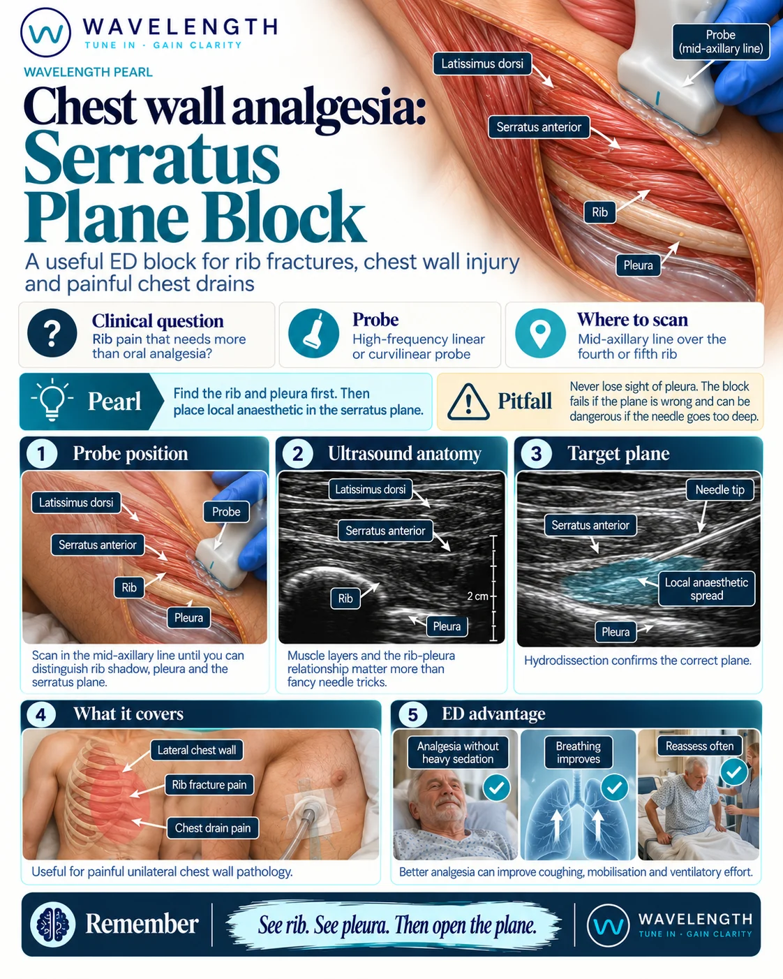
Scan images in the graphic are illustrations, not patient scans.
Before you block
- Check for a pneumothorax and consider whether a chest drain is needed.
- Explain the procedure and its risks, and record consent.
- Weigh the patient or estimate the weight, and calculate the maximum safe dose of local anaesthetic. Plane blocks use large volumes, so the dose is the main risk.
- Attach monitoring, gain IV access and know where the lipid emulsion is.
The scan
- Lie the patient supine or on the uninjured side, with the arm moved away from the chest.
- Place the linear probe in the mid-axillary line over the fourth or fifth rib, in the sagittal plane.
- Count the layers: skin, fat, latissimus dorsi at the back, serratus anterior lying on the rib, the rib with its shadow, and the bright pleura sliding between the ribs.
- Insert the needle in plane and aim at the rib, not between the ribs.
- Inject 2 to 3 mL first and watch the fluid lift the muscle away from the rib, opening the plane. Then inject the rest.
The deep approach places the injection between serratus anterior and the rib. The superficial approach places it between latissimus dorsi and serratus anterior. Both spread over the side of the chest wall.
Reading the view
- Rib. A bright, curved line with a black shadow below it.
- Pleura. A bright, shimmering line between the ribs, deeper than the rib surface.
- Correct spread. A dark, lens-shaped pool of fluid spreading along the plane, lifting the muscle.
- Wrong plane. Fluid swelling within the muscle, which looks like a bright, expanding blob.