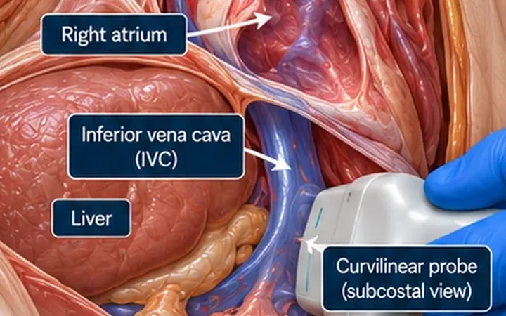
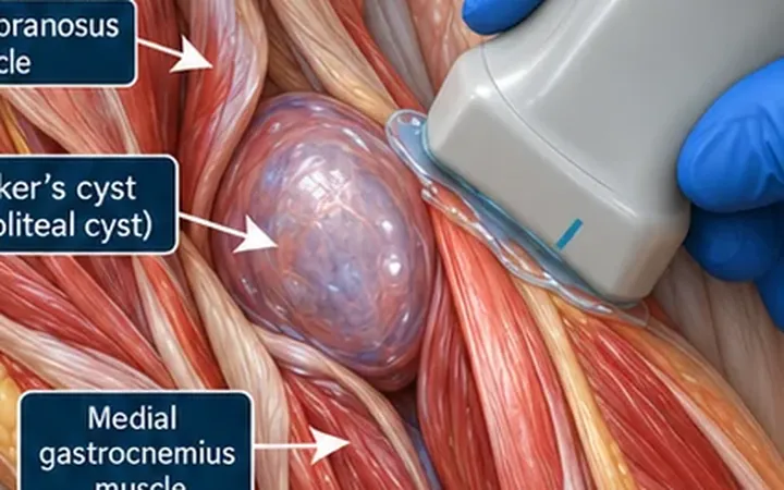
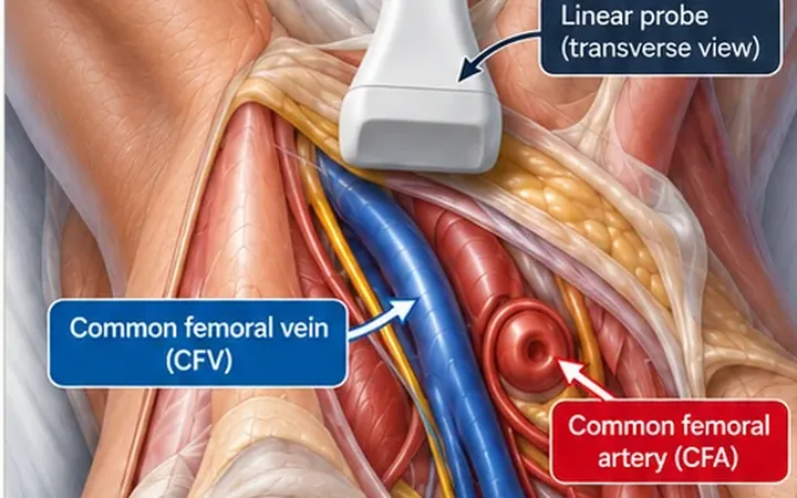
In a breathless or shocked patient, the right ventricle tells you whether the heart is under pressure from the lungs. One enlarged ventricle has many causes, acute and chronic. Combining a few findings gives a much stronger answer.
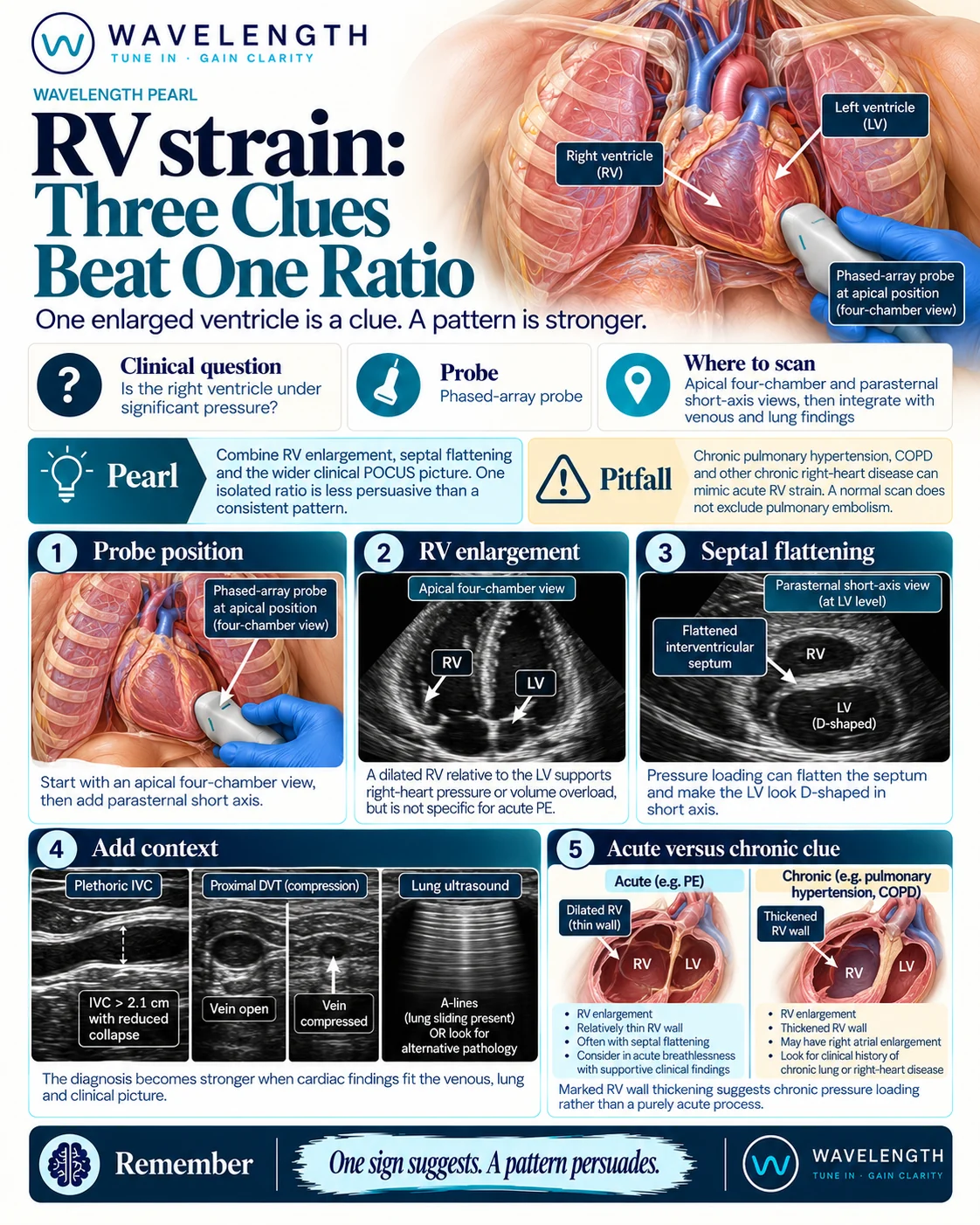
Scan images in the graphic are illustrations, not patient scans.
The scan
- Start with the apical four-chamber view using the phased-array probe. Compare the right ventricle with the left at the base.
- Move to the parasternal short-axis view at the level of the papillary muscles. Look at the shape of the left ventricle and the septum.
- Measure the right ventricular free wall in the subcostal view if you can.
- Add the IVC, compression of both legs, and the lungs.
Reading the view
- RV enlargement: in the apical four-chamber view, the right ventricle is normally about two thirds of the left. A right ventricle as wide as the left, or wider, is dilated.
- Septal flattening: in short axis, the septum flattens and the left ventricle looks like a D.
- Venous and lung clues: a plethoric IVC, a non-compressible femoral or popliteal vein, and clear lungs with A-lines all make pulmonary embolism more likely.
- Acute or chronic: a thin right ventricular wall suggests an acute rise in pressure. A free wall over 5 mm suggests chronic pressure load, such as pulmonary hypertension or chronic lung disease.
Acting on it
In a shocked patient with suspected high-risk pulmonary embolism, RV dysfunction on bedside echo supports treatment when CT is not immediately possible. Follow your local pulmonary embolism pathway and involve senior and specialist teams early.