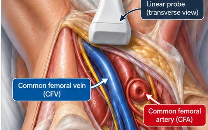
A swollen, painful calf sends you looking for a clot. When the veins compress, the answer often sits behind the knee. A Baker's cyst, or popliteal cyst, is a fluid-filled bursa connected to the knee joint. When it ruptures, fluid tracks down the calf and the leg looks exactly like a DVT. The two can also occur together.
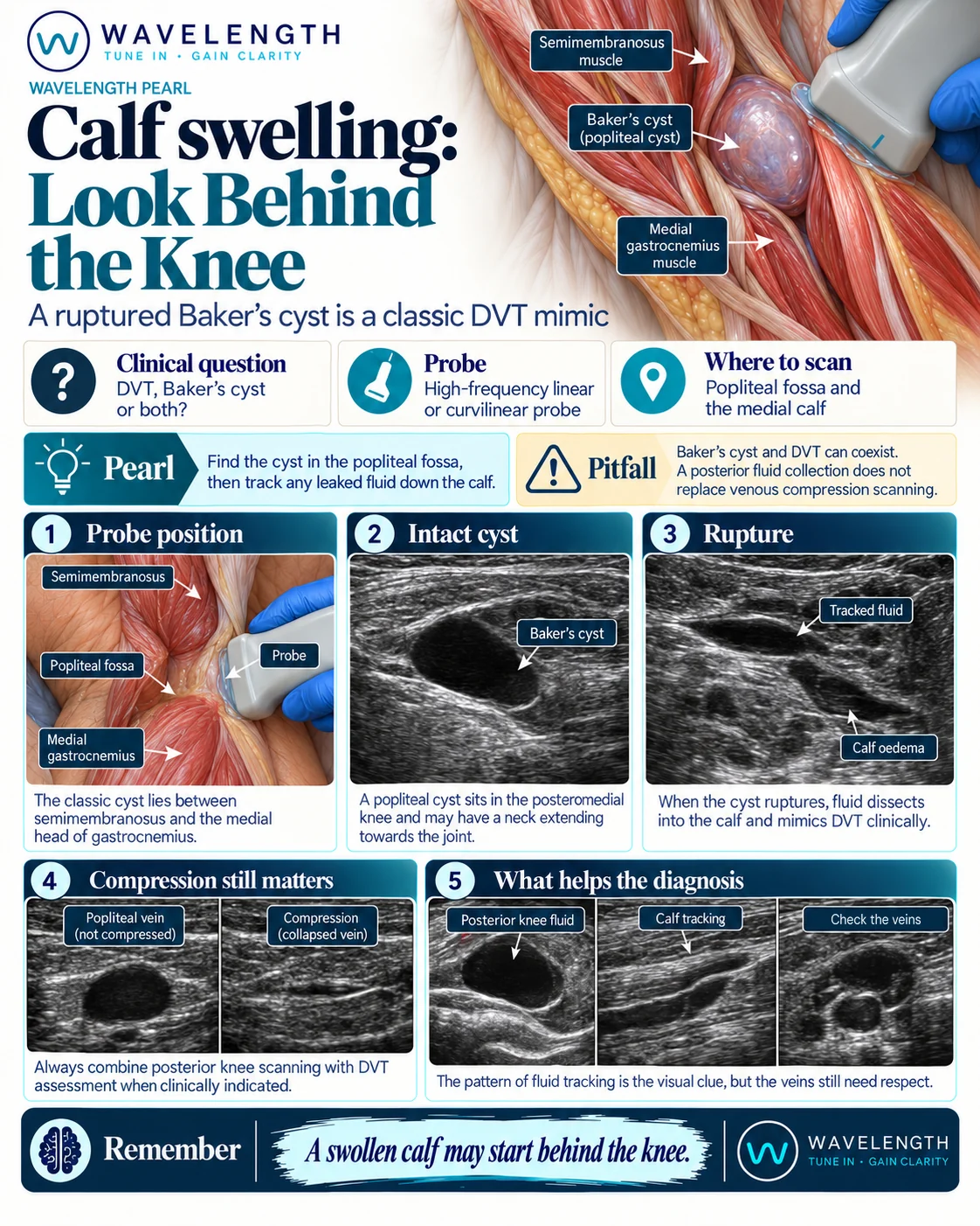
Scan images in the graphic are illustrations, not patient scans.
The scan
- Complete the proximal compression scan of the femoral and popliteal veins first, or as part of the same scan.
- With the knee slightly flexed, place the linear probe transversely over the medial side of the popliteal fossa.
- Find the medial head of gastrocnemius and the semimembranosus tendon beside it.
- Look for a collection between them, with a narrow neck running towards the joint.
- Scan the cyst in two planes and check its lower end.
- Follow the medial calf downwards, between gastrocnemius and soleus, looking for tracking fluid.
- Switch on colour Doppler over any round collection behind the knee.
Reading the view
- Intact cyst. A smooth, dark collection with a neck between semimembranosus and the medial head of gastrocnemius. It may contain debris or septations.
- Ruptured cyst. The lower end becomes pointed or irregular, and a thin layer of fluid tracks down between the calf muscles or under the skin.
- Popliteal artery aneurysm. A round structure behind the knee with flow inside on colour Doppler. It is not a cyst.