Judging left ventricular (LV) function by eye takes practice. E-point septal separation (EPSS) gives new scanners a number to anchor that judgement. It measures how close the anterior mitral leaflet comes to the interventricular septum when the valve opens in early diastole.
A well-filled, strongly contracting ventricle drives a brisk opening, and the leaflet almost touches the septum. A dilated, poorly contracting ventricle fills slowly, and the leaflet falls short.
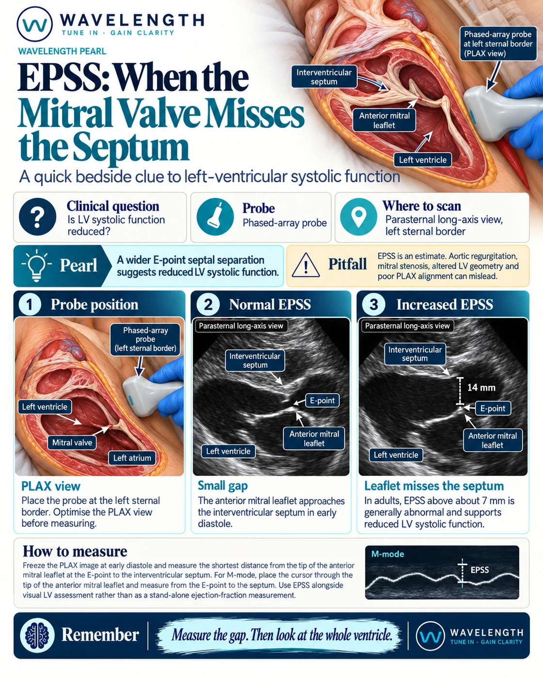
Scan images in the graphic are illustrations, not patient scans.
The scan
- Use the phased-array probe and the cardiac preset.
- Get a parasternal long-axis (PLAX) view at the left sternal border, third or fourth intercostal space.
- Optimise the view: the septum and the posterior wall lie horizontal, and the mitral valve and aortic valve both open on screen.
- Switch on M-mode and place the line through the tip of the anterior mitral leaflet.
- On the M-mode trace, the leaflet makes an M shape. The first, taller peak is the E-point.
- Measure from the E-point to the septum at that moment.
Reading the view
- Normal. The E-point comes within about 7 mm of the septum.
- Increased EPSS. A gap above about 7 mm supports reduced LV systolic function. The wider the gap, the weaker the ventricle tends to be.
- No M-mode? Freeze the PLAX loop, scroll to the frame where the leaflet opens widest in early diastole, and measure the shortest distance from the leaflet tip to the septum.
Beyond the number
- EPSS supports your visual estimate of LV function. It does not replace it, and it is not an ejection fraction.
- Check the rest of the heart: the right ventricle, the pericardium and the valves, then the lungs and the inferior vena cava.