The jugular venous pressure (JVP) tells you how full the right side of the heart is. At the bedside it is often hard to see: a large neck, a noisy department, a patient who cannot lie still. Ultrasound shows you the internal jugular vein (IJV) directly, so you see where the column of blood ends.
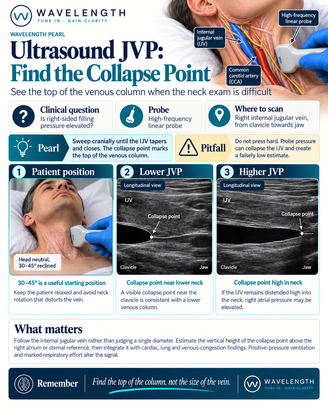
Scan images in the graphic are illustrations, not patient scans.
The scan
- Sit the patient at 30 to 45°, head supported, looking straight ahead or turned only slightly to the left. Turning the head further stretches and flattens the vein.
- Use the linear probe on the right side of the neck, in transverse.
- Use plenty of gel and almost no pressure. Let the probe rest on the gel.
- Start just above the clavicle. Find the common carotid artery (round, thick-walled, pulsatile) and the IJV beside it, lateral and usually more superficial.
- Slide the probe slowly up the neck, following the IJV.
- Stop where the vein tapers and its walls meet. This is the collapse point, the top of the venous column.
- Turn the probe to longitudinal over the vein to confirm. The vein looks like a tapering tube, closing at the top.
Reading the view
- Collapse point low in the neck, near the clavicle. The venous column is short. The right-sided filling pressure is low or normal.
- Collapse point high in the neck, or no collapse point below the jaw. The column is tall. The right-sided filling pressure is likely high.
- Estimate the height. Measure the vertical height of the collapse point above the sternal angle, as you would for a clinical JVP.
Beyond the finding
- Use the JVP with the rest of the picture: the heart, the lungs, the inferior vena cava and the patient.
- Positive-pressure ventilation, forceful breathing, severe tricuspid regurgitation and superior vena cava obstruction all change the vein and mislead the estimate.