Bedside ultrasound in suspected renal colic looks for hydronephrosis, and rules out alternatives such as an abdominal aortic aneurysm. The bladder adds one more clue. Every few seconds, peristalsis squirts urine from each ureter into the bladder. Colour Doppler shows this as a jet. A complete obstruction stops it.

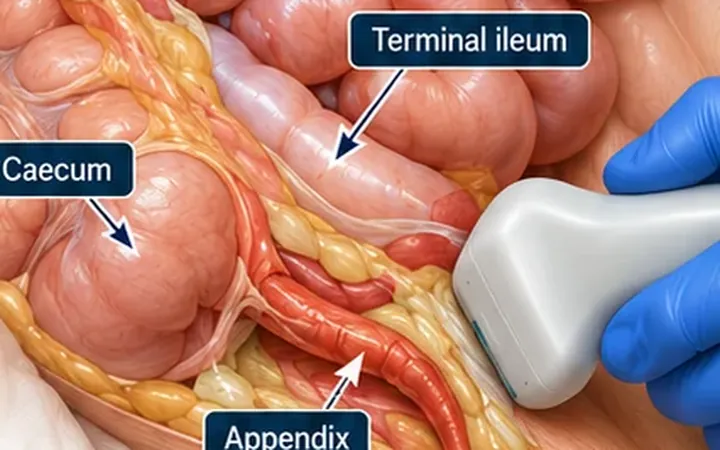
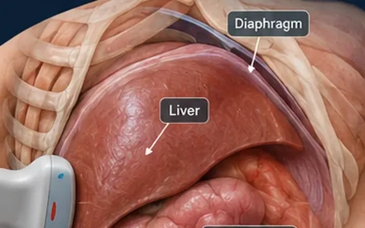
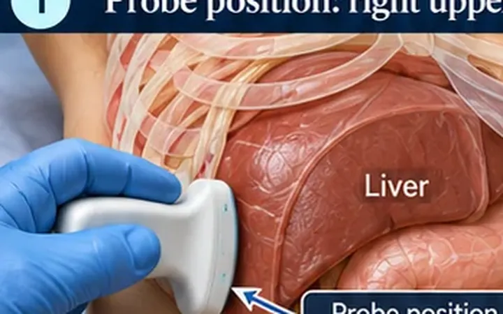
Scan images in the graphic are illustrations, not patient scans.
The scan
- Scan with a reasonably full bladder. Offer a drink if the patient can have one.
- Use the curvilinear probe in transverse above the pubic symphysis, angled down towards the base of the bladder, the trigone.
- Find the two ureteric orifices at the back of the bladder base.
- Switch on colour Doppler with the scale set low, and the box over both orifices.
- Hold still and watch for at least two to three minutes. Jets come intermittently.
- Then scan both kidneys for hydronephrosis.
Reading the view
- Normal. A flame of colour from each orifice, crossing the bladder towards the middle.
- Absent jet on the painful side, with jets on the other side, supports obstruction on that side.
- Weak jets on both sides suggest a low urine output or an underfilled bladder, not obstruction. Rehydrate and rescan.
Where it fits
In the STONE trial, starting with point-of-care ultrasound rather than CT in suspected renal colic reduced radiation exposure, with no increase in high-risk missed diagnoses. Ultrasound does not replace CT for stone size and position, or when the diagnosis is uncertain.
- A patient over 50 with first-time renal colic needs their aorta scanned.
- Fever with an obstructed kidney is an emergency. It needs urgent urology review for drainage.