The gallbladder moves with breathing, changes shape after a meal, and hides behind bowel gas. New scanners sweep the right upper quadrant hoping to see it. A landmark gets you there every time: the main lobar fissure, a bright line inside the liver which runs from the portal vein to the gallbladder neck.
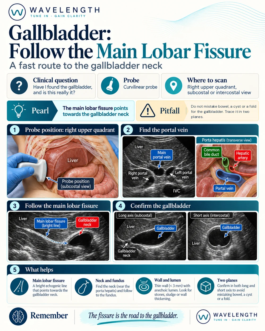
Scan images in the graphic are illustrations, not patient scans.
The scan
- Lie the patient supine. If bowel gas gets in the way, roll them onto their left side.
- Place the curvilinear probe below the right costal margin, marker to the patient's head, and ask the patient to breathe in and hold.
- Find the portal vein, a round black vessel with a bright wall at the hilum of the liver.
- Look for the bright line running from the portal vein through the liver. This is the main lobar fissure.
- Follow it. The black, pear-shaped structure at its end is the gallbladder, with its neck nearest the fissure.
- Sweep from neck to fundus in long axis, then turn 90° and sweep again in short axis.
- If the costal margin is in the way, move to the intercostal spaces in the anterior axillary line.
What to look for
- Wall. Measure the anterior wall, the one nearest the probe. Normal is under 3 mm.
- Stones. Bright, curved echoes with a black shadow behind them. Roll the patient. Stones move, polyps do not.
- Sludge. Grey, layering material without a shadow.
- Sonographic Murphy's sign. Maximal tenderness when you press the probe directly over the gallbladder.
- Pericholecystic fluid. A thin black rim around the gallbladder.
Pitfalls
- After a meal, the gallbladder contracts and its wall looks thick. Ask when the patient last ate.
- Ascites, heart failure and hypoalbuminaemia thicken the wall without cholecystitis.
- A gallbladder packed with stones shows a bright line and a dense shadow, with no black lumen. Do not mistake it for bowel gas.