Right iliac fossa pain is one of the commonest reasons for a surgical referral. Ultrasound is the first-line image in children and in pregnancy, and it avoids radiation. At the bedside, a confident positive scan speeds referral. A negative or incomplete scan leaves the question open.
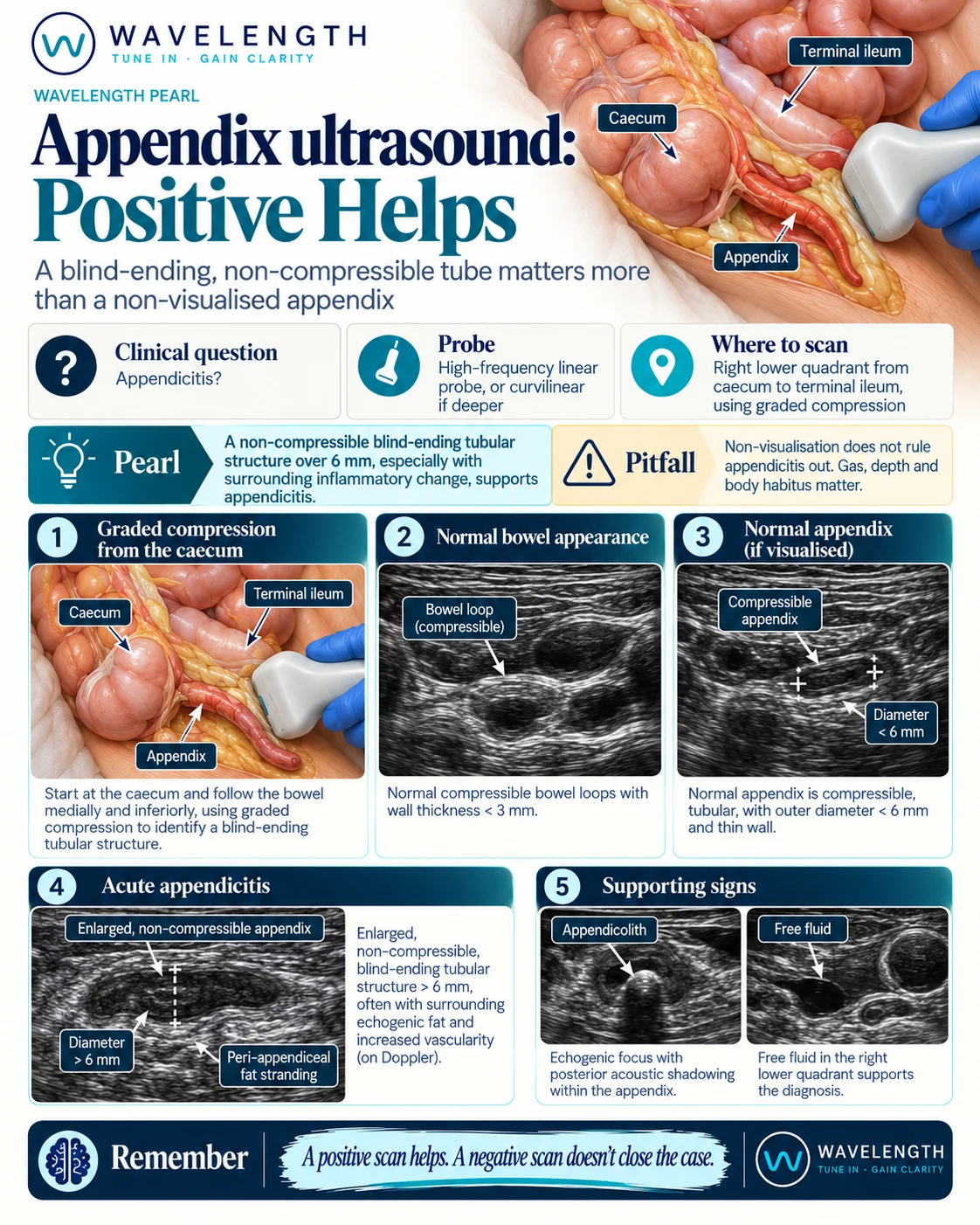
Scan images in the graphic are illustrations, not patient scans.
The scan
- Use the linear probe. Switch to the curvilinear probe in larger patients.
- Ask the patient to point to the spot where it hurts most. Start there.
- Find the caecum and the terminal ileum in transverse in the right lower quadrant.
- Press slowly and steadily with the probe, then hold. This is graded compression. It pushes bowel gas aside and shows which tubes collapse.
- Look for a tube which ends blindly, arises from the caecum, and does not compress.
- Measure its outer diameter, wall to wall, in transverse.
- Confirm in long axis.
Reading the view
- Normal bowel. Compresses under pressure and moves with peristalsis.
- Normal appendix. Compressible, thin-walled, with an outer diameter under 6 mm.
- Appendicitis. A blind-ending, non-compressible tube over 6 mm across, often with bright, inflamed fat around it and increased flow on colour Doppler.
- Supporting signs. An appendicolith, a bright focus with a shadow, inside the appendix. Free fluid or a collection in the right lower quadrant.
Acting on the result
- Positive scan: refer to the surgical team with your findings.
- Non-diagnostic scan: use the clinical picture, a scoring system and local imaging pathways. Formal ultrasound, MRI in pregnancy or CT in adults are the next steps.
- A perforated appendix is often harder to see. A collection or free fluid with a convincing history needs senior review.