A groin swelling after angiography, a femoral line or injecting drug use is a common emergency department problem. A haematoma, an abscess and a pseudoaneurysm all look alike from the outside. Treat a pseudoaneurysm as an abscess and a needle goes into an artery. Ultrasound with colour Doppler separates them at the bedside.
A pseudoaneurysm (false aneurysm) is a contained leak from a hole in the artery wall. Blood flows out through a short channel, the neck, into a sac walled by surrounding tissue, then back again.
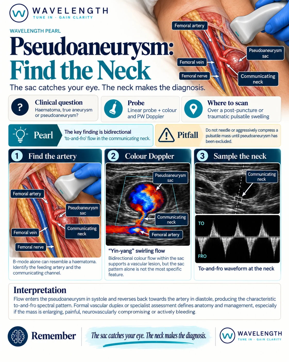
Scan images in the graphic are illustrations, not patient scans.
The scan
- Use the linear probe. Scan both groins if the history is unclear.
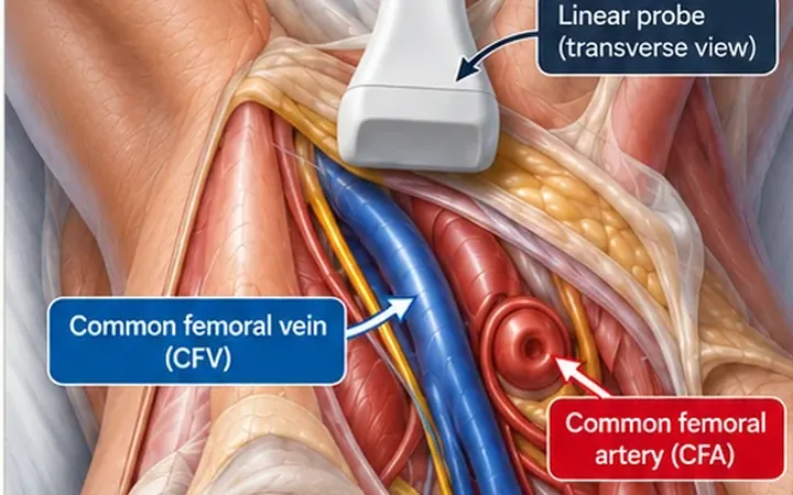
- Find the common femoral artery in transverse, then follow it to the swelling.
- Look at the swelling in B-mode. A pseudoaneurysm sac often holds layered clot and looks like a haematoma.
- Switch on colour Doppler over the swelling. Lower the scale if you see no flow.
- Look between the sac and the artery for a narrow channel carrying flow. This is the neck.
- Place the pulsed-wave Doppler gate in the neck and record the waveform.
Reading the view
- Haematoma. A collection with no flow inside it on colour Doppler.
- Pseudoaneurysm sac. Swirling flow in two directions, red and blue side by side, often called the yin-yang sign. Clot often lines part of the sac.
- The neck. A channel from the artery to the sac. Pulsed-wave Doppler shows flow into the sac in systole and back out in diastole, above and below the baseline. This is the to-and-fro waveform.
- True aneurysm. The artery itself is wider, with its own wall all the way round, and no separate neck.
Beyond the diagnosis
- Refer to the vascular or interventional radiology team. Formal duplex defines the size, the neck and the treatment.
- Urgent signs: a growing or painful mass, skin changes over it, active bleeding, or a cold or numb leg.
- Treatment options include ultrasound-guided thrombin injection, guided compression and surgical repair. Some small pseudoaneurysms close by themselves under follow-up.