Examination alone separates peritonsillar cellulitis from abscess poorly. Blind needle aspiration misses collections and risks the carotid artery, which runs behind and to the side of the tonsil. Ultrasound shows both the collection and the vessels.
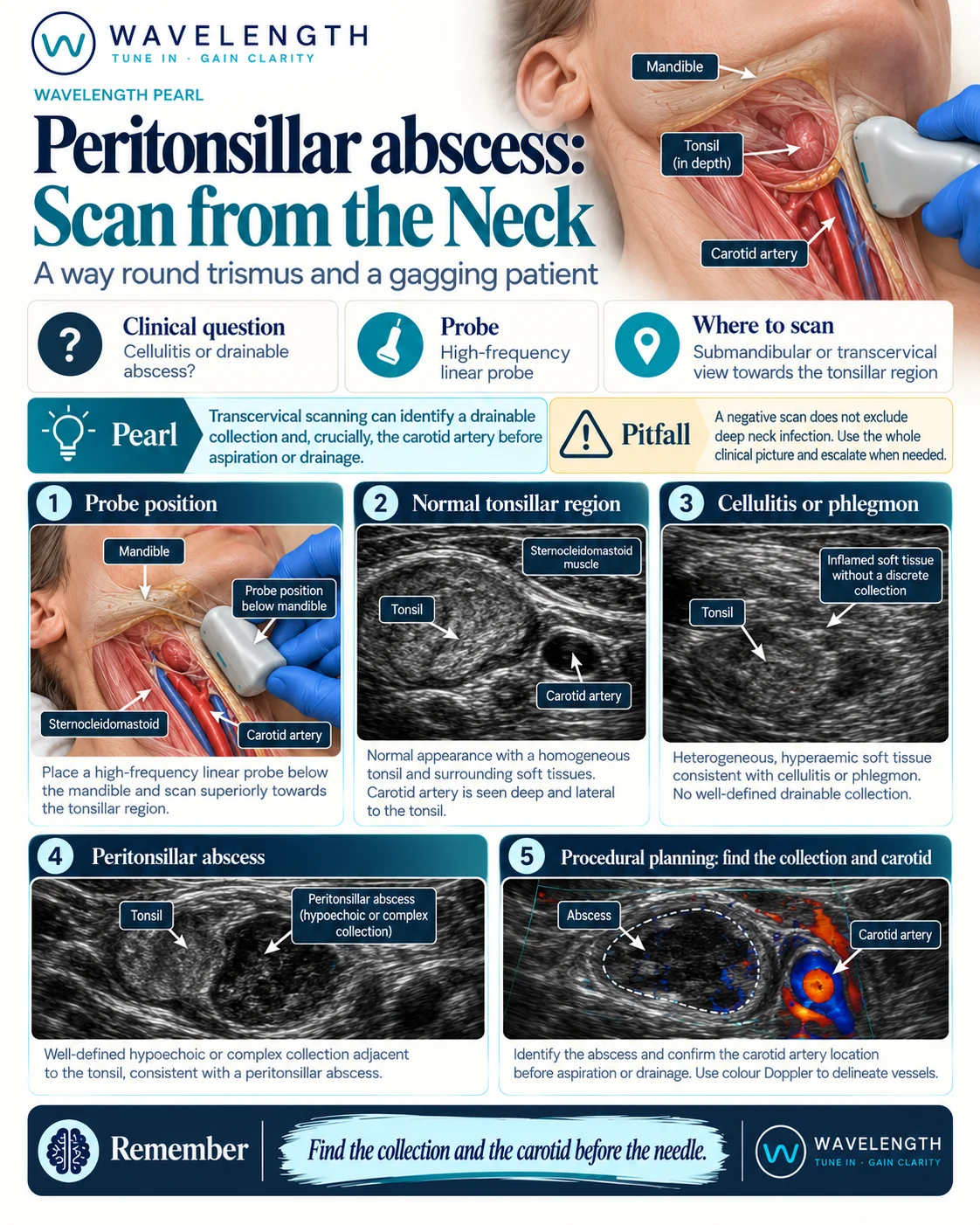
Scan images in the graphic are illustrations, not patient scans.
Two approaches
- Transcervical. A linear probe under the angle of the mandible, angled up towards the tonsil. No mouth opening needed, so it suits trismus.
- Intraoral. An endocavitary probe in a cover, placed on the tonsil. Better resolution, and it guides drainage in real time, but it needs the patient to open the mouth.
The transcervical scan
- Sit the patient up, head turned slightly away from the side you scan.
- Place the linear probe just below the angle of the mandible, then angle it up and inwards towards the tonsil.
- Find the submandibular gland and the tonsil, a grey, oval, speckled structure.
- Look for a collection beside the tonsil.
- Switch on colour Doppler and find the carotid artery, behind and to the side of the tonsil. Compare with the other side.
Reading the view
- Normal tonsil. Oval, evenly speckled, symmetrical with the other side.
- Cellulitis or phlegmon. Swollen, mixed tissue with increased flow, but no defined collection.
- Abscess. A dark or mixed collection with a defined edge beside the tonsil, with no flow inside.
Before drainage
Measure the depth of the collection and its distance from the carotid. Aim away from the artery, and do not advance the needle deeper than the collection. Follow your local ENT pathway.