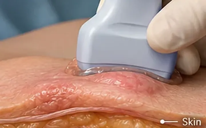
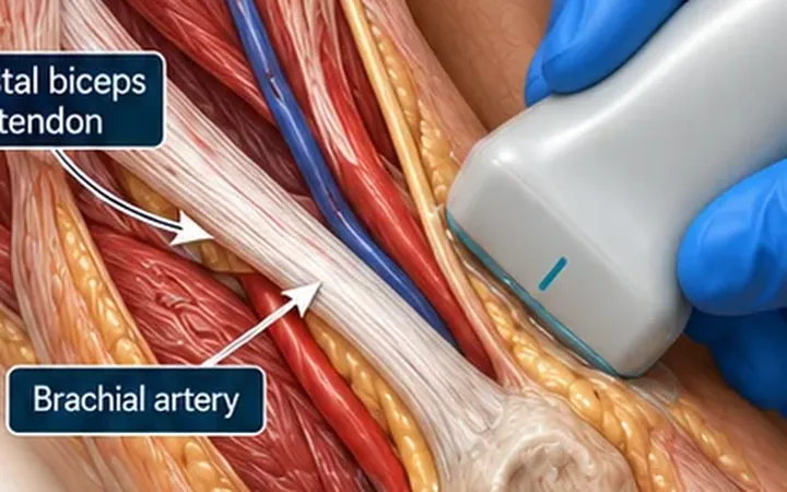
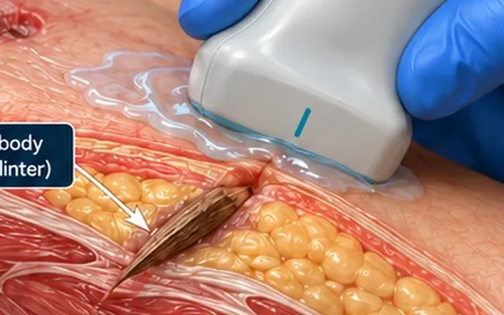
Achilles ruptures often present as a kick in the back of the heel during a sprint or a jump. Patients can still walk and plantarflex using the other calf and foot muscles, so the injury is often missed at first. The calf squeeze test is the key examination. Ultrasound adds the gap, its size and its position.
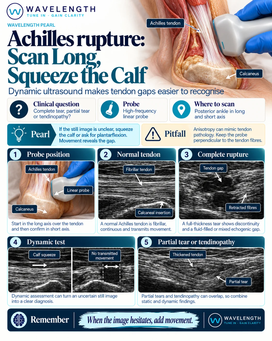
Scan images in the graphic are illustrations, not patient scans.
The scan
- Lie the patient prone with both feet hanging over the end of the bed.
- Use the high-frequency linear probe in long axis, with plenty of gel.
- Start at the insertion on the calcaneus and scan up to the calf muscles.
- Turn 90° and scan in short axis along the same length.
- Squeeze the calf, or gently plantarflex and dorsiflex the foot, while you watch the tendon in long axis.
- Compare with the other side.
Reading the view
- Normal. A continuous, bright, fibrillar band. When you squeeze the calf, the whole tendon slides as one.
- Complete rupture. A gap filled with dark fluid or mixed haematoma, often a few centimetres above the insertion. The torn ends look frayed. Fat from behind the tendon can bulge into the gap. Movement above the gap does not pass to the tendon below.
- Partial tear or tendinopathy. A thickened, darker tendon with some continuous fibres. Movement passes through.