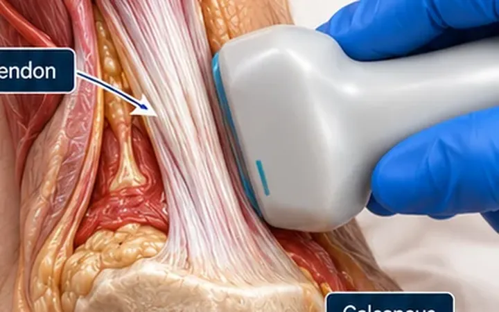
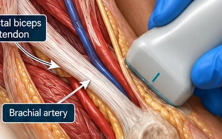
A splinter, a shard of glass or a thorn left in a wound causes pain, infection and repeat visits. Glass and metal show on X-ray. Wood, plastic and thorns usually do not. Ultrasound shows all of them, and maps their depth and their relation to tendons and vessels before you cut.
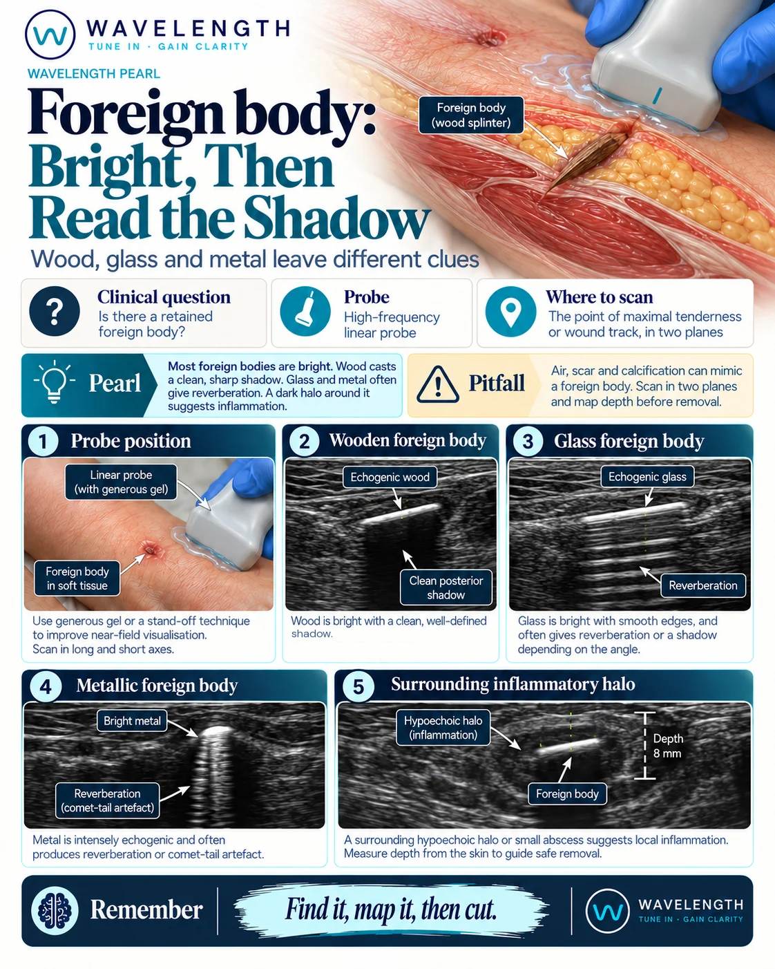
Scan images in the graphic are illustrations, not patient scans.
The scan
- Use the linear probe with plenty of gel. For fingers, use a thick layer of gel or a stand-off.
- Start at the wound and scan along the likely track, in two planes.
- Look for a bright structure. Turn the probe until it lies along its length.
- Note what lies behind it.
- Mark its position on the skin in two planes and measure its depth.
- Note nearby tendons, nerves and vessels. Use colour Doppler for vessels.
Reading the view
- Wood, plastic and thorns: a bright line with a clean dark shadow behind it.
- Glass and metal: often bright reverberation lines or a comet tail behind the object, with or without a shadow.
- A dark halo around the object suggests inflammation, granulation tissue or a small abscess. It often appears after a day or more.
Before removal
Mark the skin over the object in two planes, record its depth and the nearest structures, and consider removal under real-time guidance. Deep objects, objects near nerves, vessels or joints, and those in the hand often need specialist removal.