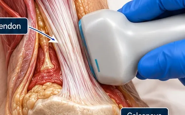
Distal biceps tears usually happen in middle-aged men lifting a heavy weight with the elbow bent. Repair works best in the first few weeks, before the tendon retracts and scars, so a missed tear costs the patient strength in supination. The examination can mislead. An intact bicipital aponeurosis, the lacertus fibrosus, holds the torn tendon close to the elbow crease, so the muscle bulge may not move far.
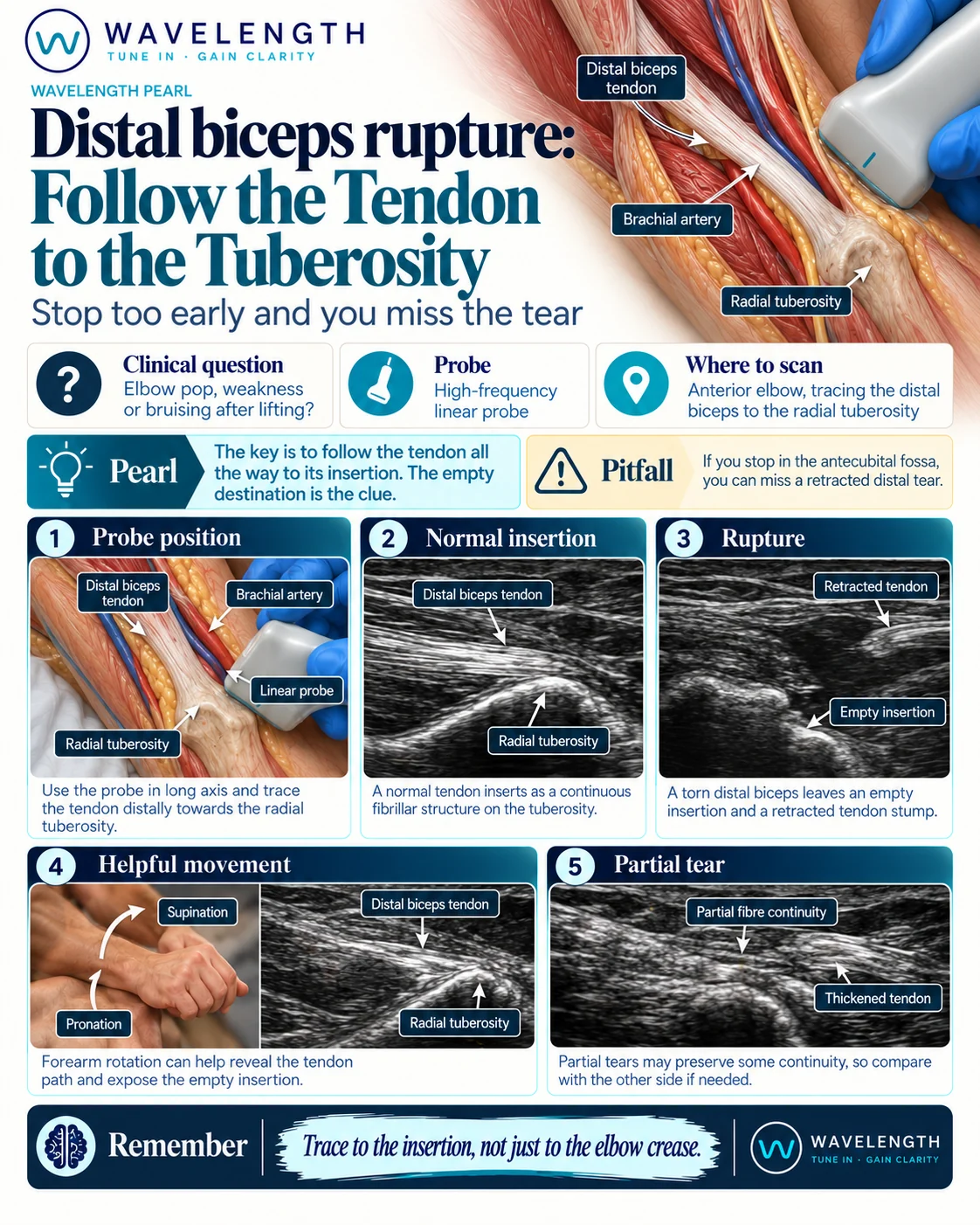
Scan images in the graphic are illustrations, not patient scans.
The scan
- Seat the patient with the elbow extended and the forearm supinated, resting on a pillow.
- Use the high-frequency linear probe, long axis, over the front of the elbow.
- Find the tendon above the elbow crease and follow it down.
- The tendon dives deep towards the radius. Press the lower end of the probe harder than the upper end, so the beam stays at 90° to the fibres.
- Follow it to the radial tuberosity, then confirm in short axis.
- Rotate the forearm slowly from supination to pronation while you watch. The tuberosity turns, and an intact tendon moves with it.
- Compare with the other arm.
Reading the view
- Normal. A continuous, bright, fibrillar tendon inserting on the radial tuberosity.
- Complete tear. An empty tuberosity, a wavy retracted stump higher up, and dark fluid or haematoma in the tendon path.
- Partial tear. A thickened or thinned tendon with dark areas but some fibres still reaching the bone.