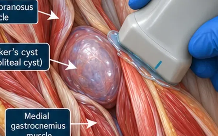
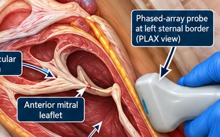
A dark stripe behind the heart looks the same whether it sits in the pericardium or the left pleural space. The difference matters. A pericardial effusion raises the question of tamponade. A pleural effusion does not. One landmark in the parasternal long-axis view separates them.
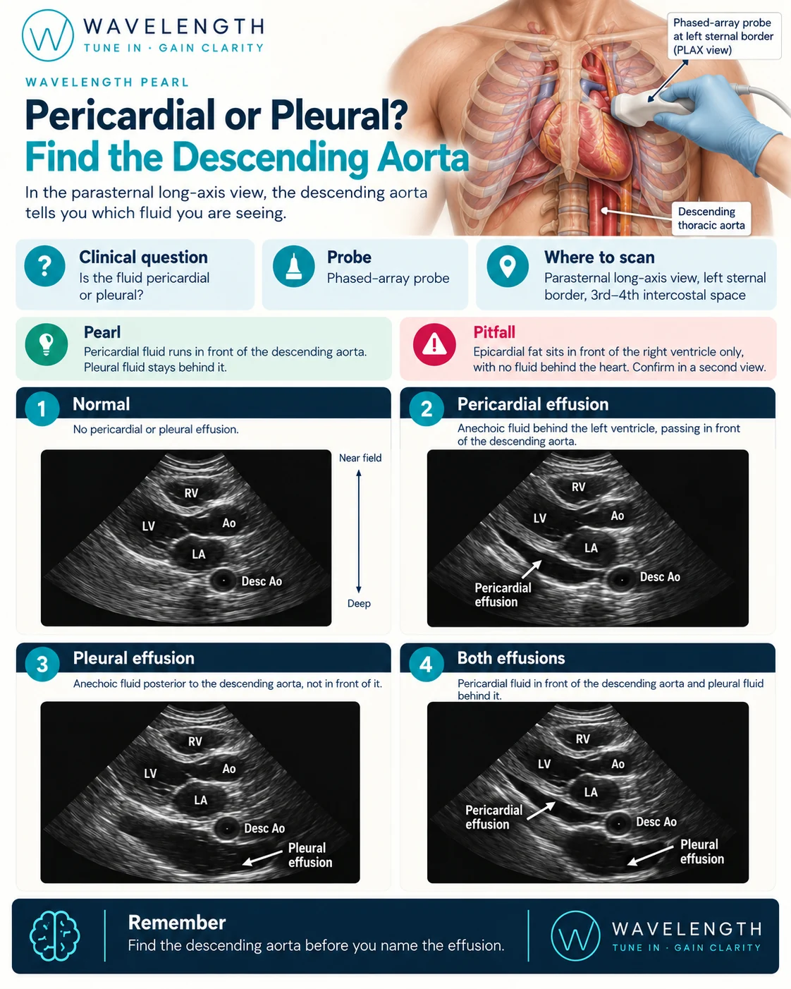
Scan images in the graphic are illustrations, not patient scans.
The scan
- Use the phased-array probe and the cardiac preset.
- Get a parasternal long-axis (PLAX) view at the left sternal border, third or fourth intercostal space.
- Increase the depth until you see well behind the heart. Fluid hides at the bottom of the screen.
- Find the descending thoracic aorta: a small round black circle in cross-section, behind the left atrium near the junction of the left atrium and left ventricle.
- Look for black fluid, and see which side of the aorta it lies.
Reading the view
- Pericardial effusion. A black stripe behind the left ventricle, between the heart and the bright pericardium. It tracks towards the left atrium and runs in front of the descending aorta, between the aorta and the heart.
- Pleural effusion. A black space behind the descending aorta. It does not pass between the aorta and the heart.
- Both. Fluid in front of and behind the aorta, separated by the bright pericardium.
Beyond the diagnosis
- If you find a pericardial effusion, look for signs of tamponade: right atrial collapse in systole, right ventricular collapse in diastole and a full, non-collapsing inferior vena cava. Tamponade is a clinical diagnosis, supported by the scan.
- If you find a pleural effusion, scan the lung bases to see its size.