Fundoscopy is difficult through a small pupil, a cataract or a bleed. The ultrasound probe looks through the closed eyelid. In a multicentre emergency department study, point-of-care ultrasound detected retinal detachment with a sensitivity of 96.9% and a specificity of 88.1%.
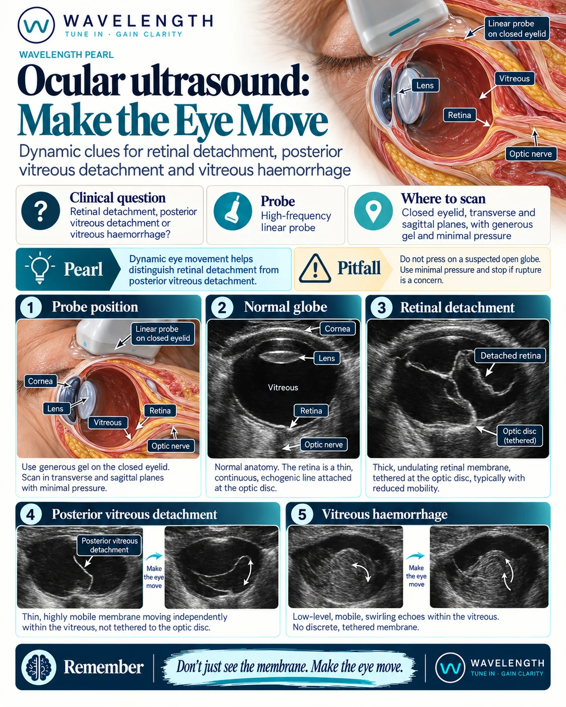
Scan images in the graphic are illustrations, not patient scans.
The scan
- Do not scan if you suspect an open globe. Pressure risks extruding the contents.
- Select the ophthalmic or ocular preset. It keeps the power within the limits set for the eye.
- Ask the patient to close the eye. Cover the eyelid with a thick layer of gel, or a clear dressing then gel.
- Rest your hand on the patient's nose or forehead so the probe floats on the gel with no pressure.
- Scan in transverse and sagittal planes through the whole globe.
- Turn up the gain to see faint membranes.
- Ask the patient to look left and right, then straight ahead, while you watch. These are the after-movements.
Reading the view
- Normal. A black vitreous, the lens at the front, and a thin bright retina lining the back of the globe, with the optic nerve shadow behind it.
- Retinal detachment. A thick, bright, often folded membrane, fixed to the optic disc at the back. It moves little with eye movement.
- Posterior vitreous detachment. A thin, faint membrane which moves freely and swings with eye movement. It does not attach to the optic disc.
- Vitreous haemorrhage. Grey, swirling echoes in the vitreous, best seen with high gain, which move like a snow globe after eye movement.