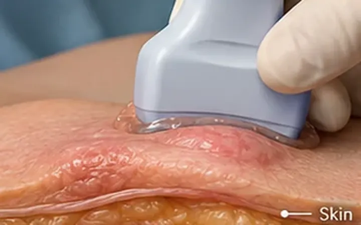
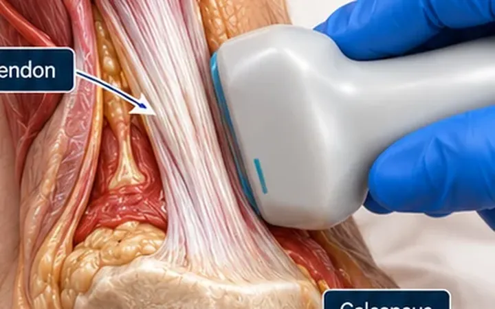
Necrotising fasciitis spreads along the deep fascia faster than the skin changes show. Early on, the leg looks like cellulitis. Pain out of proportion to the redness is often the first clue. Ultrasound looks under the skin at the bedside, while you wait for the surgeon.
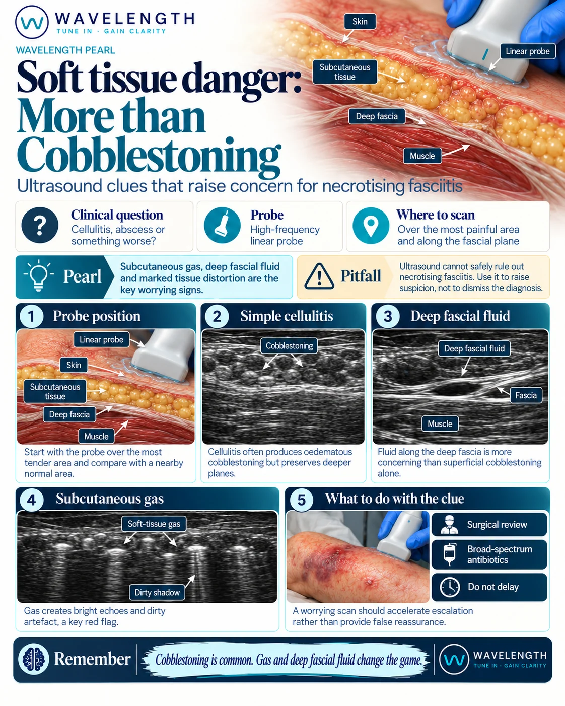
Scan images in the graphic are illustrations, not patient scans.
The scan
- Use the high-frequency linear probe. Switch to the curvilinear probe for a large thigh or buttock.
- Scan normal skin away from the redness first, to see this patient's normal fat and fascia.
- Scan the most painful area, then beyond the edge of the redness.
- Follow the deep fascia: the bright line between the fat and the muscle.
- Measure any dark fluid lying along the fascia.
- Look for bright spots in the soft tissue with grey, dirty shadowing behind them.
STAFF: what to look for
- Subcutaneous Thickening: thick, bright fat, often with cobblestoning.
- Air: bright spots with dirty shadowing or ring-down from gas in the soft tissue.
- Fascial Fluid: a dark layer of fluid along the deep fascia.
In an emergency department study, diffuse thickening of the soft tissue with more than 4 mm of fluid along the deep fascia identified necrotising fasciitis with a sensitivity of 88% and a specificity of 93%.
Reading the view
- Cellulitis. Thick, bright fat with dark strands between the fat lobules. The deep fascia stays thin and sharp, with no fluid along it.
- Worrying. Fluid tracking along the deep fascia, especially more than 4 mm, beyond the area of redness.
- Most worrying. Gas. Bright spots with dirty shadowing in the fat or along the fascia, without a wound or recent injection to explain them.